Download
1 / 41
410 likes | 531 Vues
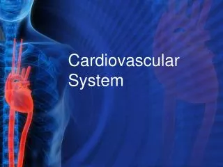
Cardiovascular System. Functions. Body’s delivery service Heart : Pumps blood through the body Blood Vessels: Carry oxygen-rich blood to all the body’s cells and return deoxygenated blood to the heart. The Heart. Average adult heart- fist-sized

E N D
Functions • Body’s delivery service • Heart: • Pumps blood through the body • Blood Vessels: • Carry oxygen-rich blood to all the body’s cells and return deoxygenated blood to the heart
The Heart • Average adult heart- fist-sized • Lies in thoracic cavity, between the lungs • Two-thirds of the heart actually lies to the left of the sternum • Amazingly powerful muscle • Beats an average of 72 times per minute, 100,000 times a day and 3000 million times (3 trillion) in the average lifetime
Parts of the Heart • The Pericardium: • A protective sac that covers the heart • Has two layers: • Visceral pericardium: innermost layer next to the heart • Parietal pericardium: outer layer
Parts of the Heart • The Heart: • Three layers of tissue: • Epicardium: outer layer • Myocardium: middle layer, muscular tissue • Endocardium: inner layer, forms membrane lining the chambers and valves
The Heart • The heart is divided into right and left sides • Each side has two chambers • Right Side: • Right Atrium: upper chamber • Right Ventricle: lower chamber • Left Side: same as right • Two sides are separated from each other by a partition called the septum
The Heart • Valves of the Heart: • Control blood flow to and from the heart • Valves that control blood between atria and ventricles: • Bicuspid (mitral) valve: Valve between left atrium and ventricle • Tricuspid valve: Between right atrium and ventricle • Valves that control blood leaving the heart (preventing backflow): Pulmonary and Aortic Valves
Blood Flow Blood Flow Animation • Blood flows through the chambers of the heart in only one direction • The flow is regulated by the valves of the heart • Arteries: carry blood AWAY from the heart • Usually carry blood rich in oxygen • EXCEPTION: Pulmonary Artery, which carries blood low in oxygen AWAY from the heart, toward the LUNGS to pick up OXYGEN from the ALVEOLI
Blood Flow • Veins: carry blood TOWARD the heart • Usually carry blood low in oxygen • EXCEPTION: Pulmonary Veins, which returns blood that is rich in oxygen TOWARD the heart. • This blood is coming from the lungs
Cardiac Conduction System • The heart muscle works like a pump. • Electrical impulses control the pumping cycle of the heart muscle. • Electrical impulses begin in one part of the heart and travel all through the heart. • The special tissues in the heart that produce electrical impulses form the cardiac conduction system
Cardiac Conduction System • The Cardiac Conduction System contains: • The Sinoatrial (SA) node • The Atrioventricluar (AV) node • Bundle of His (AV Bundle) • Bundle Branches • Purkinje Fibers (network)
SA Node • Small round structure • Located in upper part of right atrium • Natural pacemaker-makes the heart start to beat • Fires at about 60-100 times per minute • Fire: transmit a nervous impulse or electrical signal • Conduction begins in the SA node (which means that each heartbeat begins here as well)
AV Node • Also small and round structure • Located along the floor of the right atrium • Receives impulses traveling through the heart (from the SA node) and can delay or slow down the impulse • If SA node is not working, AV node can also act as the pacemaker • Usually fires at a rate of 40-60 times per minute
Bundle of His • Located next to the AV node • Transfers electrical energy from the atria to the ventricles • When impulses reach the ventricles, they are divided into the bundle branches
Bundle Branches • Located along the left and right side of the septum separating the left and right ventricles • Impulses travel through the left and right bundle branches to the left and right ventricles • Fork in the Road: Some impulses go right, others left • Make the heart muscle contract
Cardiac Conduction Animation!! Purkinje Network • Spreads the impulses throughout the ventricles, through a system of fibers called the Purkinje fibers • Fibers provide an electrical pathway for each of the cardiac cells • Electrical impulses speed up here • Activate right and left ventricles at one time, causing them to contract • Electrical impulse produces an electrical wave, which can be recorded using an Electrocardiogram (ECG)
Circulation • There are three types of circulation: • Coronary • Pulmonary • Systemic
Coronary Circulation • Circulation of blood within the heart • Coronary Arteries: branch off the aorta to supply blood to the heart muscle • AORTA: main artery through which blood exits the heart • Fun Fact: the heart needs more oxygen to function than any other organ (except the brain)
Pulmonary Circulation • Flow of blood between the heart and the lungs • Pulmonary arteries: carry blood LOW in oxygen from the right ventricle of the heart to the lungs, where it will pick up oxygen from the alveoli • Blood RICH in oxygen flows back to the left atriumthrough vessels called the Pulmonary veins
Systemic Circulation • Flow of blood between the heart and the cells of the body • The heart pumps blood through the arteries to the cells of the body • The muscular contraction of the heart pushes the blood through the arteries • Blood going from the heart to the cells of the body is RICH in oxygen
Special Arteries • Specialized arteries are those that carry oxygen-rich blood to specific areas of the body • Carotid Artery: supplies blood to the head and neck • Femoral artery: supplies blood to the thigh • Arterioles: smaller divisions of arteries • Capillaries: smallest blood vessel • Deliver nutrients and oxygen to cells • Remove waste products from cells
Systemic Circulation • After flowing through the capillaries, blood begins the trip back to the heart through venules, which are small veins • The Veins take blood that is LOW in oxygen back to the heart
Systemic Circulation • The blood from the upper part of the body is collected and returns to the heart through the Superior Vena Cava • Blood from the lower part of the body collects and returns to the heart through the Inferior Vena Cava • Both vessels return blood to the Right Atrium Animation!!!
Diseases of the Cardiovascular System • Hypertension • Stroke • Aneurysm • Coronary Artery Disease (CAD) • Myocardial Infarction (MI, Heart Attack) • Congestive Heart Failure (CHF)
Hypertension: high blood pressure • Blood Pressure: the force of the blood surging against the walls of the arteries • No symptoms • Usually the result of lifestyle factors • Medication is available to keep high blood pressure at a normal level, but it needs to be taken every day • Untreated, it may cause major damage to the liver, blood vessels and kidneys • Can lead to a cerebrovascular accident (CVA) or stroke
Stroke • Can occur when a blood clot blocks the flow of blood in a vessel, or when a blood vessel bursts in the brain
Stroke • May experience mild symptoms before a major stroke • Sudden numbness or weakness of the face, arm or leg especially on one side of the body • Facial droop • Sudden confusion • Difficulty speaking or understanding • Trouble seeing in one or both eyes • Sudden difficulty in walking, loss of balance, coordination or dizziness • Sudden, severe headache
Stroke • Short incidents of blood flow loss (transient ischemic attacks-TIAs) • Can recover completely from a mild stroke • Severe stroke damage may be permanent Face Arms & Legs Speech Time
Stroke • Common damage from a stroke • Thought disorders • Speech difficulty • Loss of muscle control • Some paralysis • Disorientation Stroke Animation!!
Arteriosclerosis • Hardening of the arteries • Arteries lose elasticity (their ability to stretch) and their ability to contract • Commonly occurs as a result of aging • Can lead to high blood pressure or an aneurysm
Aneurysm • Occurs when a disease or defect at birth weakens the walls of an artery • May also be the result of trauma • Trauma: a physical injury caused by external force or violence • Ballooning out of an artery wall • Common sites: • Aorta • Abdominal artery • Cerebral artery
Aneurysm • May cause pain and pressure, but usually there are no symptoms • If found before it ruptures (pops), most must be surgically removed • If they do rupture, they can cause severe bleeding and death
Coronary Artery Disease (CAD) • Narrowing of the coronary arteries that supply blood to the heart • Usually caused by atherosclerosis, which is a buildup of fatty plaque inside the blood vessels • Can lead to angina or myocardial infarction
Myocardial Infarction (MI) • Also known as a heart attack • Blood flow to the heart is cut off • Can result in permanent damage to the heart tissue • Without a supply of fresh blood, deprived heart tissue begins to die • If a large portion of the heart is affected , patient may die
Myocardial Infarction (MI) • Angina: temporary loss of blood supply to the heart • May cause same symptoms as heart attack • Symptoms of a Heart Attack: • Chest discomfort, feeling of fullness in the chest, or pain or pressure in the center of the chest that lasts for more than 3-5 minutes • Light-headedness or dizziness • Sweating or breaking out in a cold sweat • Fainting • Nausea, vomiting • Shortness of breath • Pale or grayish skin color • Generalized weakness
Congestive Heart Failure (CHF) • Occurs when heart cannot pump at its usual capacity • Vital organs do not receive enough blood • May occur as a result of atherosclerosis and/or MI
Congestive Heart Failure (CHF) • Heart is weakened and cannot pump at its usual force • If the right side of the heart is weakened, blood backs up in the organs and builds up • If left side of heart is weakened, blood builds up in lung tissue CHF Animation!!


