Heart Sounds
250 likes | 382 Vues
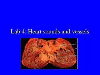
Heart Sounds. 2 distinct sounds associated with closing of valves 1 st - AV valves open in response to atrial contraction, blood flows through, they close 2 nd - SL valves snap shut after ventricular contraction Heart murmurs- abnormal heart sounds Obstructions in blood flow

Heart Sounds
E N D
Presentation Transcript
Heart Sounds • 2 distinct sounds associated with closing of valves • 1st- AV valves open in response to atrial contraction, blood flows through, they close • 2nd- SL valves snap shut after ventricular contraction • Heart murmurs- abnormal heart sounds • Obstructions in blood flow • Thin walls vibrating due to blood flow • Valve problems
Cardiac Cycle • Events associated with blood flow through the heart during one complete heartbeat • Systole- contraction, diastole- relaxation • Atrial systole & diastole followed by ventricular systole & diastole • Mechanical events follow events of EKG • Ventricular filling: mid-to-late diastole • Ventricular systole: ventricles begin to contract, isovolumetric contraction phase, expel blood • Isovolumetric relaxation: early diastole
Cardiac Output • Amount of blood pumped by each ventricle in 1 min. • Cardiac Ouput= Heart Rate x Stroke Volume • Stroke volume- V of blood pumped out per ventricle per beat • Ave CO= 75beats/min x 70mL/beat= 5.25L/min
Abnormalities and Disorders • Angina pectoris- thoracic pain from deficiency of blood to myocardium • Myocardial infarction- “heart attack”- prolonged coronary blockage- cells die, replaced by noncontractile scar tissue • Atheroscleorsis- patchy thickenings “plaques” intrude vessels • Congestive heart failure- CHF- pumping efficiency of heart is so low that blood circulation is inadequate to meet tissue needs • progressively worsening condition
Irregularities and Disorders • If SA node becomes nonfunctional other slower “pacemakers” dominate contraction speed • Slower impulse through myocardium allows some muscle fibers to contract well before others, resulting in reduced pumping effectiveness • Arrhythmias- irregular heart rhythms • Fibrillation- condition of rapid, irregular out-of-phase contraction in heart, rhythm is taken away from the SA node by rapid activity in other heart regions • Heart block- interferences with ability of ventricles to receive impulses- damage to AV node • If no impulses get through (total heart block), ventricles beat at own pace, far too slow to maintain adequate circulation • “Pacemakers” are used to recouple the activities of the atria and ventricles
http://image.slidesharecdn.com/ar-141204080734-conversion-gate01/95/blood-vessels-arteries-veins-and-capillaries-2-638.jpg?cb=1417680755http://image.slidesharecdn.com/ar-141204080734-conversion-gate01/95/blood-vessels-arteries-veins-and-capillaries-2-638.jpg?cb=1417680755
Vascular System • Arteries- blood away from heart • Run deep, well protected by body tissues • Veins- blood to heart • Superficial & deep, deep run with arteries • Capillaries- exchange of materials (diffusion) • 3 layers (tunics) of walls of blood vessels • Tunica intima- innermost, endothelium, reduce friction of blood flowing through lumen • Tunica media- middle, mostly circular smooth muscle & sheets of elastin • Arteries- vasoconstriction & vasodilation • Tunica externa- outmost- loosely woven collagen fibers that protect & reinforce
Blood Vessels- Arterial System • Elastic (conducting) arteries- thick-wall, near heart, largest & most elastic (most elastin) • Conduct blood from heart to medium-sized arteries • Muscular (distributing) arteries- deliver blood to specific body organs • Vasoconstriction & vasodilation • Arterioles- smallest arteries • little more than 1 layer of smooth muscle tissue around endothelium- go into capillary beds
Blood Vessels- Capillaries and Venous System • Capillaries- microscopic vessels, tunica intima only, very porous • Venus System • Venules- form when capillaries unite, all endothelium, very porous, largest have 1-2 layers of smooth muscle cells & thin externa • Veins- 3 distinct tunics, thinner walls with larger lumens than arteries
Blood Flow, Pressure and Resistance • Blood flow- V of blood flowing through vessel/ organ/entire circulation, in a given period (mL/min) • Blood Pressure (BP)- force per unit area exerted on vessel wall by contained blood (mmHg) • Generally refer to systemic arterial pressure in largest arteries near heart • P gradient keeps blood moving from areas of high to low P • (Peripheral) Resistance- opposition to flow, measure of friction blood encounters as it passes through vessels, 3 sources: • Blood viscosity (thickness/stickiness of blood), blood vessel length, blood vessel diameter • Relationship between flow, pressure & resistance: F= ∆P/R
Systemic Blood Pressure • Arterial pressure- 2 factors- elasticity of elastic arteries & V of blood forced into them at any time • Systolic P= ave 120mmHg • Diastolic P= ave 70-80mmHg (enough P to continue flow into smaller vessels) • Difference between systole & diastole Ps= pulse P • Capillary Blood Pressure- low is better for fragile, extreme permeable walls • Venous Blood Pressure- too low for adequate return, needs help! • Respiratory pump, muscular pump & smooth muscle (sympathetic nervous system) • Valves and wide lumen
Maintaining Blood Pressure • Cooperation of heart, blood vessels & kidneys (under control of brain/hormones) • Factors: cardiac output (CO), peripheral resistance (PR) & blood volume (BV) ∆P= CO x R (CO= F) • CO also affected by venous return • Short term controls alter R & CO • Blood vessel diameter & distribution of blood
Vasomotor Center and Input from Higher Brain Centers • Medulla contains vasomotor center- oversees changes in diameter of blood vessels • Sympathetic caused overall vasoconstriction & BP (& vice versa) • Higher Brain Centers- cerebral cortex & hypothalamus can modify arterial Ps via relays to medulla • exp- hypothalamus- fight-or-flight response
Vasomotor Center and Input from Baroreceptors and Chemoreceptors • Receptors located in major arteries (internal carotid artery (to brain), aortic arch & most thoracic & cervical arteries) • Baroreceptors- respond to changes in arterial P/stretch • Inhibit vasomotor center vasodilation (RP, venous reservoirs, CO) • Rapid response, protect circulation from short-term, acute changes in BP • Chemoreceptors- respond to changes in levels of blood O2, CO2, & H+ • if pH s or CO2s, impulses to cardioacceleratory center (to ↑CO) & to vasomotor center (vasoconstriction)
Arteries Brachiocephalic trunk R and L Common carotids R and L Subclavians Axillary Brachial Decending aorta (thoracic and abdominal) Renal External and internal iliac Femoral Saphenous Veins Brachiocephalic External and intenral jugulars Subclavian Axillary Brachial Long thoracic vein- blood from chest muscles and lungs Adrenolumbar Renal veins Common iliac veins External and internal iliac Femoral Great saphenous Major/Specific Blood Vessels • Note thymus and phrenic nerve • Label: • Aorta (arch) • Coronary arteries • Vena cavae (sup and inf, in cat= precava and postcava) • Pulmonary trunk • Pulmonary arteries • Pulmonary veins
Blood Facts • Varies in color from scarlet (O2 rich) to dark red (O2poor) • More dense & about 5x as viscous as H2O • pH 7.35-7.45 • Temp 38˚C (100.4˚F) • 8% of body weight (ave V= 5-6L males, 4-5L females) • Connective tissue, has cellular & liquid components
Functions of Blood • Distribution- carries O2 & nutrients to, & picks up metabolic wastes (CO2, NH3, etc) from, all cells in body, transports hormones • Regulation- maintain body temp, pH, & adequate fluid V in circ sys • Protection- prevents own loss (clotting) & infection
Plasma • 55% of blood volume • Straw colored sticky fluid, 90% water • 8% plasma proteins by V • Albumin- (60%) main protein contributor to osmotic P (Na+ other), shuttles other molecules through circulation, acts as buffer • Globulins- (36%) mostly transport lipids, metal ions, & fat soluble vitamins, some also act as antibodies in immune response • Fibrinogen- (4%) forms fibrin threads of blood clot
Formed Elements • 45% of blood volume • Erythrocytes~ 45%= hematocrit • Leukocytes & platelets ‹1%= buffy coat • Odd features- • 2 of 3 aren’t true cells • Most survive only a few days in blood stream • Most do not divide (made from cell division in bone marrow) • Hematopoiesis- blood cell formation by hematopoietic stem cells in red bone marrow
Platelets • Platelets- cell fragments • essential for clotting process occurring in plasma when vessel or lining is ruptured
Hemostasis- Stoppage of Bleeding • Fast, localized, carefully controlled, complex series of reactions • 3 steps: • Vascular spasms- immediate response (vasoconstriction) • Injury to smooth muscle chem..s released by endothelial cells & platelets, reflexes initiated by nociceptors • Platelet plug- temporarily seals break • Stick to collagen fibers of damaged endothelial lining • Once activated, release chemicals • exp. Serotonin- enhances vascular spasm • Coagulation (blood clotting)- blood transformed from liquid to gel, 3 phases: • 1) prothrombin activator forms • 2) converts prothrombin (plasma protein) to thrombin (enzyme) • 3) thrombin catalyzes joining of fibrinogen molecules into a fibrin mesh • Over 30 substances involved!- procoagulants, anticoagulants
Disorders in Hemostasis • Thrombus- clot in unbroken blood vessel • Embolus- thrombus breaks away for the wall & floats freely in the blood stream • Embolism- embolus becomes trapped
Erythrocytes- RBCs • Carry O2 & CO2, hemoglobin (Hb)- protein easily & reversibly binds with gasses • Oxyhemoglobin or deoxyhemoglobin • Mature have no nuclei or organelles • No mito= don’t use O2 • Life span ~100-120 days • Trapped by circulatory channels (especially spleen), fragmented, engulfed by macrophages, destroyed • Blood doping- athletes draw off RBCs & inject a few days before an event, s O2 carrying capacity • Anemia- abnormally low O2 carrying capacity • Insufficient #RBCs, low hemoglobin content, abnormal hemoglobin
Blood Transfusions and Blood groups • Whole blood transfusion- routine when blood loss is rapid & substantial • Packed RBCs (most plasma removed)- restore O2 carrying capacity • Human blood groups- RBC plasma membranes have highly specific glycoproteins (antigens) at external surfaces • If transfused blood is recognized as foreign transfused cells may become agglutinated (clumped together) & destroyed by immune cells • Agglutinogens- RBC antigens