Download

1 / 32
460 likes | 2.7k Vues
UCLA Head & Neck Surgery Resident Lecture Series Marc Cohen, M.D. Laryngeal complications of endotracheal intubation. A little history…. Video tutorial. A little history…. A little history…. By 1910, intubation for anesthesia had become an accepted practice

E N D
UCLA Head & Neck Surgery Resident Lecture Series Marc Cohen, M.D. Laryngeal complications of endotracheal intubation
A little history… • By 1910, intubation for anesthesia had become an accepted practice • During WWI, Magill and Macintosh made profound improvements • In 1970, high-volume, low pressure cuffs were introduced
Prolonged intubation vs. tracheotomy? • In the 1960’s, long term intubation for the management of premature LBW infants was recommended • Until…. Subglottic stenosis was recognized
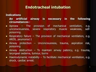
Indications for endotracheal intubation • 1. Temporary relief of upper airway obstruction • 2. Assisted ventilation for respiratory failure • 3. Pulmonary toilet
What are the potential complications of endotracheal intubation? • Edema • Granuloma • Healed fibrous nodule • Interarytenoid adhesion • Posterior glotticstenosis • Subglotticstenosis • Healed furrows • Ductal cysts • Hematoma • Laceration • Subluxation of arytenoid cartilage • Loss of mobility of cricoarytenoid joint • Vocal cord paralysis • Nasogastric tube syndrome
Pathogenesis Pressure-Induced Injuries • Vulnerable structures • Medial surfaces of arytenoids • Vocal processes • Cricoarytenoid joints • Cricoid cartilage • Posterior glottic/Interarytenoid region
Pathogenesis • Supraglottic structures may become edematous, but rarely sustain serious damage • Tracheal injuries have also become less significant due to low pressure cuffs • Although there is potential for injury if the cuff is inflated too high
Pathogenesis • The microcirculation of the mucosa and mucoperichondrium is interrupted when pressure from the ETT exceeds capillary pressure • Ischemia Necrosis Edema, Hyperemia, Ulceration, and Erosion
Factors for susceptibility • Extrinsic factors • Diameter of ETT • Duration of intubation • Traumatic or multiple intubations • Patient factors • Poor tissue perfusion (i.e. sepsis, organ failure, etc) • LPR • Abnormal larynx • Wound healing, keloid • Movement • During ventilator use • During suctioning • During coughing • During transport
“Laryngeal Bedsore” • Superficial ulceration can occur within hours of intubation • Usually heals without scarring • As ETT pressure continues, migration of inflammatory cells ensues • If epithelial erosions are incomplete, epithelium may be replaced by squamous metaplasia • Further pressure causes ulceration through mucosa to cartilage • Causes perichondritis and destructive chondritis • As opposed to superficial damage, deeper ulceration heals by secondary intention and fibrosis
Edema • 3 locations • Reinke’s space Usually persists after extubation • Ventricular mucosa, seen as “protrusion” Usually resolves after extubation • Subglottis Usually resolves after extubation
Granulation tissue • Seen within 48 hours • Proliferate at periphery of ulcerated areas
Granulation tissue • Flaps of granulation tissue • Can move with inspiration/expiration • Inspiratory stridor • Not recommended to excise both sides • Most cases will resolve without any intervention once ETT is removed
Granulation tissue • Incomplete resolution of granulation tissue can yield: • Postintubation granuloma • Healed fibrous nodule
Posterior glottic stenosis • Forms when scar contracts after wide ulceration with no intact median strip of mucosa • Vocal cords unable to abduct • Glottis remains partly closed • Inspiratory stridor • Voice is usually unaffected • Treatment: deep vertical division with laser or 11 blade down to level of cricoid • Re-stenosis is likely • Costal cartilage graft may be necessary (endoscopically or open)
Subglottic stenosis • Many causes • In infants, most common factors related to acquired SS are ETT size and LPR during long-term intubation • Presentation in an infant: • Failed extubation • Recurrent or atypical croup • Slowly progressive airway obstruction • Difficulty passing ETT • Postanesthesia stridor
Cotton-Myer Grading System • Grade I - < 50 % obstruction • Grade II – 51-70% obstruction • Grade III – 71-99% obstruction • Grade IV – No detectable lumen • Rule of thumb: • Subglottic diameter < 4.0 mm in a full-term infant is the lower limit of normal (< 3.0 mm in a preterm infant)
Subglottic stenosis • When repeated attempts at extubation fail: • Reintubate with smaller ETT • Racemic epinepherine • Dexamethasone • If these maneuvers fail: • Cricoid split with/without cartilage graft • Tracheostomy
Ductal Cysts • Result from retention of mucus in obstructed, dilated ducts of submucosal mucous glands • Most are small and require no treatment • When large and cause obstruction, endoscopic removal is required
Arytenoid dislocation • May occur during passage of an ETT • Left arytenoid is usually affected since intubation occurs from right side of mouth • Patient will complain of hoarseness, throat discomfort, odynophagia, and cough • Microlaryngoscopy and closed reduction should be performed early
Nasogastric tube syndrome • Occurs when NGT rests centrally, rather than laterally • Anterior wall of hypopharynx/posterior wall of cricoid becomes ulcerated • Results in perichondritis, chondritis, necrosis • Can progress to sudden, life-threatening bilateral vocal cord paralysis due to myositis of PCA muscles • Diabetics and renal transplants who are in renal failure are especially vulnerable • Warning signs: hoarseness, otalgia, and odynophagia • Treatment: remove NGT, abx, G-tube, and possible tracheostomy
Timeline of postextubation obstruction • Immediate: flaps of granulation tissue, laryngeal spasm • Minutes to hours: flaps of granulation tissue, subglottic edema, granulation tissue, LPR • Days to weeks: persistent edema or granulation tissue, granuloma • Months: posterior glottic stenosis, subglottic stenosis
To trach or not to trach? • One school of thought is that anyone who is intubated longer than 7 days should undergo tracheotomy • Newer recommendations are for DL after 7 days – if no evidence of significant laryngeal pathology, keep the patient intubated unless plan for long-term tracheostomy