Download
1 / 43
440 likes | 942 Vues
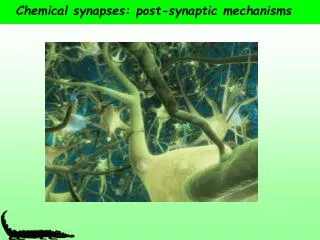
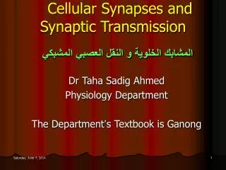
Cellular Synapses and Synaptic Transmission المشابك الخلوية و النقل العصبي المشبكي. Dr Taha Sadig Ahmed Physiology Department The Department ’ s Textbook is Ganong. Q :What are neural synapses ? المشابك العصبية They are areas of contact between neurons .

E N D
Cellular Synapses and Synaptic Transmissionالمشابك الخلوية و النقل العصبي المشبكي Dr Taha Sadig Ahmed Physiology Department The Department’s Textbook is Ganong
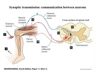
Q :What are neural synapses ? المشابك العصبية They are areas of contact between neurons . They are essenial for communication between different neural components of the nervous system Neuromuscular synapses are areas of contact between nerve and muscle ( neuromuscular junctions )
Sensory or motor information is transmitted for long or short distances by berves in the form of action potentials (APs ) . • When the information is needed to be passed from one neuron to another or from a motor nerve to a muscle , a synapse is needed . • Each bit of information as it is passed from one neuron to another may be (1) blocked , or (2) passed , to be added to other bits of information reaching that neuron . • In this way synaptic transmission permits grading and adjustment of neuronal activity .
Classification of Synapses According to Type • (A) Chemical synapses : Employing neurotransmitters ( chemical transmitters ) that is secreted by the presynaptic nerve to combine with receptors on the membrane of the postsynaptic cell . • These are the ones that coomonly occur in the CNS & NMJ and concern us in this lecture . • (B) Electrical Synapses : Here the membranes of the presynaptic & postsynaptic cells come very close together , forming a low-resistance bridge for transmission of electrical current between the two cells . • These will not be discussed in this lecture .
Classification of Synapses on the Basis of Their Location • (1) Axo-dendritic • (2) Axo-axonal ( axo-axonic ) • (3) Axo-somatic , • & less commonly • (4) Dendro somatic • (5) Somato-somatic • NB : Soma means cell-body
Some neurons have , only few synapses on them , but others may have as many as 10,000 synapses on their soma and dendrites.
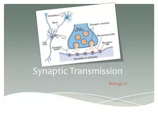
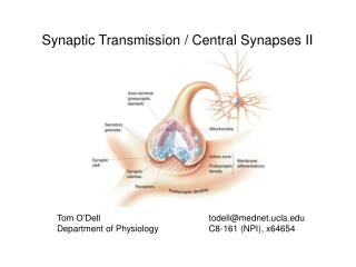
How ? When a nerve Terminal or Axon Terminal ( presynaptic cell ) makes contact with the cell-body or dendrites of another ( postsynaptic ) cell . The end of the presynaptic terminal may enlarge , forming a synaptic knob Postsynaptic Membrane Synaptic knob Presynaptic Cell Presynaptic Membrane Postsynaptic Membrane Postsynaptic Cell Neuron )
Synaptic knobتضخّم فينهاية الخلية السابقة للمشبك What are the components of a synapse ? الخلية السابقة للمشبك Presynaptic Cell • Synaptic knob ( axon terminal , nerve terminal ),of pre-synaptic cell contains the Neurotransmitter, e.g Acetylcholine • Synaptic cleft الأخدود المشبكي , a microscopic space , contains the enzyme which destroys the Neurotransmitters e.g., Cholinesterase • Post-synaptic membrane which contains the Receptors الخلية التالية للمشبك Postsynaptic Cell
Neurotransmitters ( Chemical Transmitters )الناقلات الكيميائية • الناقلات الكيميائية هي مواد ترسلها الخلية ما قبل المشبك Presynapticcell ثم تعبر الأخدود المشبكي Synaptic Cleft لتتحد مع المستقبلات في الخلية التالية للمشبك Postsynaptic Cell لتحفزها أو لتحبطها ، حسب نوع الناقل العصبي ( لو كان تحفيزي أو إحباطي ) • Neurotransmitters are chemical substances released by some neurons ( called Presynaptic cell ) , cross the synaptic cleft , and bind to receptors on the dendrites or soma of other neurons ( the Postsynaptic cell ) .
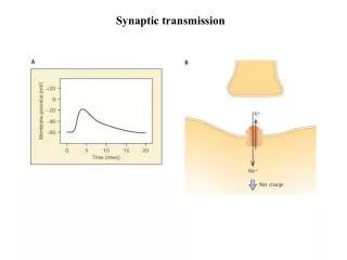
Neurotransmitters can be Excitatory تحفيزية/ محفزة or Inhibitory for Synaptic Transmission تثبيطية/ مثبطة/ محبطة للنقل المشبكي • Excitatory Neurotransmitterالناقل المحفّزة( التحفيزي) : Is a transmitter that produces Excitatory Postsynaptic Potential ( EPSP)تحفيز محلّي on the postsynaptic neuron . • The EPSP is a Local Responseتحفيز كهربائي محلي ، لا ينتقل بعيدا that decreases the membrane potential of the postsynaptic cell , bringing it closer to the Firing (Threshold) Potential عتبة إنطلاق الآكشن بوتنشيال • In other words , the EPSP is a local depolarizing potential that makes the Postsynaptic Cell more liable (easier) to fire ( produce ) Action Potentials و يالتالي جاعلا إياها مشحونة و أكثر قابلية للأستثارة و بالتالي أكثر قابلية و جاهزية لأطلاق آكشن بوتنشيال
Excitatory Neurotransmitter (Contd) Q: Is the EPSP an ALL-OR-None response ? Q : Is it proportional to the strength of the stimulus ? Q : Can several EPSPs sum( add up ) ? • Yes , if several EPSPs on the post-synaptic cell sum ( by the process of summation ) they may bring the membrane to its threshold ( firing ) level and then an AP will be fired by the post-synaptic cell.
Examples of excitatory transmitters : • (1) Acetylcholine : Opens sodium channels in the Postsynaptic Cell Membrane depolarization EPSP . • (2) Glutamate : Produces EPSP by opening of calcium channels Long – Term Potentiation (LTP) الأستثارة طويلة الأمد • Glutamate Receptors play important role in a process Of long-term synaptic facilitation which is called Long – Term Potentiation (LTP) . • This LTP is a is essential for formation of memories in the brain
Inhibitory Neurotransmitter الناقل المثبط و المحبط • when it combines to its receptors , it produce Inhibitory Postsynaptic potential (IPSP) that hyperpolarizes the post-synaptic cell , thereby making it less excitable ( more difficult to excite , more difficult to produce APs ) . • Examples of inhibitory transmitter is • GABA ( which in some places opens chloride channels , and in others opens potassium channels ). • Enkephalin : Inhibitory transmitter . Found in the GIT and spinal cord . In the spinal cord it exerts analgesic activity مخفف للشعور باللم , reducing the feeling of pain .
Q: What happens to the transmitter after it has combined with its postsynaptic receptors and produced it effect ? In the synaptic cleft there are enzymes that will then destroy the receptor : In case of : • Acetylcholine Acetylcholinesterase (Ach-esterase) ; • Noradrenaline ( Norepnephrine ) it is COMT (Catechol-O-Methyl Transferase )
Recycling of Vesicles in the Presynaptic Membrane Q : Where is the neurotransmitter synthesized ( made) ? In the Cell-Body Q: Where does filling of vesicles with the transmitter occurs ? In the nerve terminal ( nerve –ending )
Q: What are the factors that affect synaptic transmission ? : Alkalosis makes the synapses more active Hypoxia and Acidosis depress synaptic transmission .
PROPERTIES OF SYNAPTIC TRANSMISSION 1/ ONE WAY CONDUCTION Why ? 2/ SYNAPTIC DELAY الزمن المستغرق بين التحفيز و ظهور الأستجابة .. ما سبب هذا التأخير ؟ The time taken between stimulation of the pre-synaptic ending and elicitation of the post-synaptic response . المتوسط عبر كل ساينابس لحاله هو 0.5 millisecond Average synaptic delay in one synapse=0.5 ms (0.5 – 0.6 ms). • Therefore , in 2 synapses = 1.0 – 1.2 ms, • in 3 synapses = 1.5 – 1.8 ms , • in 4 synapses = 2.0 – 2.4 ms , etc
3/ Divergence النشر و التضخيم & 4/ Convergenceالأجتماع في هذه الشبكة العصبية الآكسون ب ينقسم diverges ليعطي الخليتين أكس و واي و من ناحية أخري : فرعان من الأكسونين أ و ب يجتمعان converge علي تزويد الخلية أكس • Advantages of divergence (1) spread of information . (2) amplification of the post-synaptic responses . Example : in the sympathetic system one pre-ganglionic neuron can innervate up to 20 post-ganglionic neurons
4/ CONVERGENCE • Example : the spinal motor neuron (anterior horn cell , AHC ) receives between 1000 – 10,000 synaptic inputs : Some of these terminals are excitatory ( produce EPSPs ). • And others are inhibitory( produce IPSPs ) on the soma or dendrite of the post-synaptic cell. • Advantage : Helps in (1) Spatial Summation , (2)integration and modulation of information .
Summation : Spatial & Temporal • Spatial summation التحفيز بالجماعة : due to adding up of EPSPs produced by more than one synaptic knob . Thus activity in one synaptic knob facilitates activity in another. • Temporal summationالتحفيز بالألحاح ، حتي ولو كان من فرد : Repeated afferent stimuli ( even if from a single synaptic knob ) cause new EPSPs before previous EPSPs have decayed.
5/ SPATIAL SUMMATION • This is a form of increased excitability of the post-synaptic membrane induced by several pre-synaptic terminals acting on several areas of the cell-membrane but firing at the same time : • The synchronous firing of these pre-synaptic terminals induces multiple EPSPs at different parts of the membrane of the post-synaptic cell these EPSPs add up ( spatiallly ) moving the membrane potential value to the firing ( threshold ) level & thereby inducing a post-synaptic response .
5/ Spatial Summation (Facilitation) (1) • A and B converge on X , • B diverges on X and Y. • A stimulus applied to either of A or B will set up an EPSPin X. • If A and B fire at the same time the two EPSPs produced by them simultaneously in two areas of X will sum ( spatial summation ) & their resultant will make X reach threshold and fire . In this case X is said to be in the discharge zoneof the afferent volley , and the EPSPS produced on it by A and B are said to facilitate each other .
Next let us consider neuron Y . It has not fired , but its excitability has been increased by the EPSP produced on it by B . Such neurons in which the excitability has been increased by an afferent but has not yet reached the firing level are said to be in the subliminal fringeمنطقة الأثارة التي لم تصل بعد حد إطلاق آكشن بوتنشيال , and it will be easier for an AP in neuron C to make Y fire . Spatial Summation (Facilitation) (2)
6/ TEMPORAL SUMMATION • This is a form of increased excitability of the post-synaptic membrane induced by a single pre-synaptic terminal : • Repetitive firing of the pre-synaptic terminal produces successive EPSPs that add up quickly in time ( temporally summate ) moving the membrane potential value to the firing ( threshold ) level & thereby inducing a post-synaptic response .
7/ Occlusion • If B fires rapidly & repetitively repetitive EPSPs will be produced n X and Y , before previous ones have decayed & will add up by Temporal Summation , and both X and Y will fire . • If C fires repetitively Y and Z will similarly discharge bt temporal summation . • However , if B and C fire repetitively at the same time X, Y and Z will discharge . • Thus the response to repetitive firing of B and C together ( 1+1=3) is less than the response to repetitive stimulation of B and C separately , because both B & C end on neuron Y ( which is common between them ) • This decrease in the expected response du e to presynaptic fibers sharing postsynaptic neurons is called OCCLUSION
8/ INHIBITION A/ Presynaptic Inhibition An inhibitory neuron , not acting directly on the target cell , but makes axo-axonal synapse on an excitatory ending that ends on the target cell . This inhibitory interneuron releases GABA which acts via either : • (1) GABAa receptors that increase chloride conductance decreasing calcium entry into the excitatory synaptic knob reducedor absent vesicle’ release ; or BABA may act via (2) GABAb receptors which , through G-protein increase potassium conductance , thereby decreasing calcium entry into the synaptic knob of the excitatory neuron.
B/Postsynaptic Inhibition ( also called Direct Inhibition ) The inhibitory interneuron acts directly on the target cell ( and not indirectly upon a cell that acts on the target cell , compare this diagram with the diagram in the previous slide ). Example of this Direct Inhibition is Reciprocal Inhibition in the spinal cord , which occurs by means Reciprovcal Innervation الأحباط المتبادل Activity in spindle afferent , besides exciting the motoneuron supplying the agonist muscle , activates an inhibitory interneuron that directly inhibit the motoneuron supplying the antagonist muscle .
C/Feedback Inhibition ( Renshaw Cell Inhibition ) • Neurons may also inhibit themselves in a negative feedback fashion ( Negative Feedback inhibition ). • A spinal motoneuron gives a collateral that synapses on an inhibitory interneuron. • This inhibitory interneuron is called Renshaw cell. This Renshaw cell, in turn , sends back inhibitory axons that terminate on the cell body of the original motoneuron as well as other motoneurons . • These axons secrete an inhibitory transmitter that produces IPSPs on cell-bodies of motoneurons and inhibit them .
D/ Feed-forward Inhibition • Example : In the cerebellum Basket and Stellate cells are excited by Granule cells via the parallel fibers and their output inhibits the Purkinje cells .
E/ Lateral ( Surround ) Inhibition • The best example of this is in the visual cortex منطقة البصر في الدماغ Activation of a particular neural sensory unit is associated with Inhibition of sensation of surrounding nearby units ( it sends collateral fibers to inhibit its neighbours ) يسكت من هو حوله ليتكلم هو