Download
1 / 32
330 likes | 379 Vues
Discover the intricate organization and functions of brain lobes, the cerebrum, cortex structure, and brainstem in vertebrates. Delve into the neural intricacies that enable quick responses in animals. Learn about CNS, PNS, and lobes.

E N D
Nervous SystemChapter 48, 49Campbell & Reece 2 Principle Regulatory Systems: Endocrine & Nervous Nervous System: gives animals capacity for quick response (endocrine hormones act more slowly, long term)
THE EVOLUTION OF THE NERVOUS SYSTEM Comparisons: simple-->complex • many invertebrates have neurons forming a diffuse network; primitive brains (ganglia) for responding to stimulus • Vertebrates: nervous system is dorsal, enclosed and protected by bones of vertebral column and skull • Trend is toward increased cephalization of control in the brain
ORGANIZATION OF THE VERTEBRATE NERVOUS SYSTEMThe Nerve Cell, "neuron":
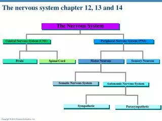
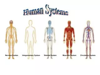
(1) CNS= spinal cord and brain • (2) PNS = (peripheral nerves): all other nerves (branches off CNS) • (a) sensory (afferent) NS: carries impulses to CNS • (b) motor (efferent) NS: carries impulses from the CNS, typically to muscles and glands • somatic NS- controls skeleton muscle (voluntary) • autonomic NS- controls smooth and cardiac (involuntary) muscles and glands • sympathetic NS- stimulate • parasympathetic NS- inhibit organizational chart
The Brain • Adult brain weighs approx. 1.4 kg (~3 lbs) and contains about 100 billion neurons • capable of carrying out many simultaneous operations • made of multiple lobes, parts
The CEREBRUM • the largest portion of the human brain (80% of brain by volume) • it is here that things like perception, imagination, thought, judgment, and decision occur • divided into right and left halves (hemispheres), connected by a tightly-packed mass of nerve fibers, the corpus callosum • "Cross-wired": right side of brain receives messages and sends signals to left side of body • Left / Right Hemispheres: responsible for different functions; • examples: injury to left side of brain results in speech impairment (aphasia); right side = musical talent... • white matter: myelinated axons, inner portion of cerebrum • grey matter: unmyelinated axons, make up outer (cortex) region of cerebrum
Cerebrum, continued • Internal structure- 3 hollow bulges- "ventricles" filled with Cerebrospinal Fluid • The wrinkly, folded surface of the cerebrum -- the cerebral cortex -- is about 2 to 4 mm thick • The wrinkles (convolutions) have "ridges" which are called gyri (singular: gyrus), and "valleys" which are called sulci (singular: sulcus).
Some of the sulci are quite pronounced and long, and serve as convenient boundaries between four areas of the cerebrum called lobes.
The furthest forward is the frontal lobe. It seems to be particularly important: • this lobe is responsible for voluntary movement and planning • is thought to be the most significant lobe for personality and intelligence • At the back portion of the frontal lobe, along the sulcus that separates it from the parietal lobe, is an area called the motor cortex. • In studies with brain surgery patients, stimulating areas of the motor cortex with tiny electrical probes caused movements. • It has been possible for researchers to actually map out the motor cortex quite precisely. • The lowest portions of the motor cortex, closest to the temples, control the muscles of the mouth and face. • The portions of the motor cortex near the top of the head control the legs and feet.
Behind the frontal lobe is the parietal lobe It includes an area called the somatosensory cortex, just behind the sulcus separating this lobe from the frontal lobe. • Again, doctors stimulating points of this area found their patients describing sensations of being touched at various parts of their bodies. • Just like the motor cortex, the somatosensory cortex can be mapped, with the mouth and face closest to the temples and the legs and feet at the top of the head.
At the side of the head is the temporal lobe The special area of the temporal lobe is the auditory cortex. • As the name says, this area is intimately connected with the ears and specializes in hearing. • It is located near to the temporal lobe's connections with the parietal and frontal lobes.
At the back of the head is the occipital lobe • At the very back of the occipital lobe is the visual cortex, which receives information from the eyes and specializes, of course, in vision. • The areas of the lobes that are not specialized are called “association cortex”. • Besides connecting the various sensory and motor cortices, this is also believed to be where our thought processes occur and many of our memories are ultimately stored.
The Brainstem • Spinal cord is continuous to the brainstem • "Brainstem" controls muscles and glands in head, autonomic regulatory functioning (resp., B.P.)
Upper Brainstem (see figure 50-4, pp. 1006) "Diencephalon" • Thalamus: • 2 egg-shaped orbs; • main relay center between brainstem and cerebrum • processes sensory information • Hypothalamus: • just below thalamus; • thermostat for mammals • maintenance of homeostasis • source of several releasing hormones The Limbic System: includes parts of the thalamus, hypothalamus, and deep cerebrum. Responsible for EMOTION, MOTIVATION, MEMORY, and DRIVES (sex, hunger, thirst, pain, pleasure, anger). Emotions are translated into actions.
Lower Brainstem: extension of the spinal cord • midbrain: mainly for relay for visual and auditory information; reflex center • pons: relay center between cerebral hemispheres and the cerebrum and cerebellum • medulla oblongata: controls autonomic homeostatic activities, like heartbeat, respiration, etc. • reticular formation: • controls consciousness, arousal • filters incoming stimuli and discriminates the "important" from the "unimportant".
TheCerebellum • The cerebellum is involved in the coordination of voluntary motor movement, balance and equilibrium and muscle tone. • It is located just above the brain stem and toward the back of the brain. It is relatively well protected from trauma compared to the frontal and temporal lobes and brain stem. Cerebellar injury results in movements that are slow and uncoordinated. Swaying and staggering when walking, and sometimes slurred speech can occur when damaged.
PROTECTION OF THE BRAIN (pp. 1007) • surrounded by three protective layers, called MENINGES. • dura mater (outermost) • arachnoid layer • pia mater (innermost) • also cushioned by a clear fluid, CEREBROSPINAL FLUID, which also fills the cavities (ventricles) within the brain.
THE SPINAL CORD (pp. 1008) • a long column of nerves that run down the center of the vertebral column • outer sheath of white matter (mostly axons), surrounding inner core of grey matter (cell bodies of neurons) • 31 pairs of spinal nerves branch outward, carrying messages to/from PNS
Transmission of Nerve Impulses • Neurons: the functional units of the Nervous System surrounded and insulated by the other nerve cells • Axons and Dendrites = "nerve fibers" • dendrites pick up/generate nerve impulses, then pass it through the cell body, and down the axon. • Myelin sheath: fatty insulation. In the PNS, this insulating material is produced by cells called Schwann cells. • insulator, speeds up impulses (100 m/sec, 225 mph) • gaps in the myelin sheath are called the nodes of Ranvier. • impulse jumps from node to node
TYPES OF NEURONS: • Sensory neurons: initiate a nerve impulse when triggered by a stimulus (dendrites embedded in sense organs) • Interneurons: (association neurons) relay nerves, transfer impulses to/from PNS to/from CNS ("connectors") • Motor neurons: axon ends in a muscle or gland. These trigger a response to a stimulus.
Synapses • junctions across which neurons communicate with each other (electrical/chemical) • impulse must be carried across synaptic cleft by a "chemical bridge" (neurotransmitter) ex: acetylcholine or noradrenalin • synapse animation • Neurotransmitters are made in cell body and are stored in synaptic vesicles at end of nerve fiber • Ca 2+ allows neurotransmitter to be released into synaptic cleft, and attaches to receptor sites on adjacent nerve fibers • acetylcholine is quickly broken down with enzyme (cholinesterase)
THE NERVE IMPULSE • The Ionic Basis of Action Potential • At rest, [K+] greater inside nerve fiber; [Na+] greater outside nerve fiber. • Resting Potential- slight excess of negative charge inside membrane from outward diffusion of K+ • Action Potential- portion of membrane becomes momentarily permeable to Na+ ions (flow inward), accompanied by an outward flow of K+
action potential animationanimation of ion flow through gated channels"Sodium-Potassium Pump": reverses (resets) polarity (at nodes) Na/K Pump animation
The Senses • Sensory organs (eyes, ears, skin, etc.) allow us to detect, and then respond to stimulus • Survival depends on evaluating and reacting to changing situations around us.
The Senses TYPES OF RECEPTORS: • Mechanoreceptors: touch, position, hearing • Chemoreceptors: taste and smell • Photoreceptors: vision, light • Thermoreceptors: heat These are all nerve endings (dendrites) embedded in body parts, sense organsA different part of the brain is responsible for interpretation of each sense, for example, the temporal lobe of the cerebrum is the hearing/sound center...
The Ear • detection of sounds - hearing. • detection of movement and the position, of the head - balance. Anatomy of the Ear animation 1. Human ear has three divisions: an outer, middle, and inner ear. 2. Outer ear consists of pinna (external flap) and the auditory canal. a. Auditory canal opening is lined by fine hairs that filter air. b. Modified sweat glands in auditory canal secrete earwax to guard ear against foreign matter. 3. Middle ear begins at tympanic membrane and ends at a bony wall with membrane-covered openings (oval window and round window). a. It contains ossicles: malleus (hammer), incus (anvil), and stapes (stirrup). b. Malleus adheres to tympanum; stapes touches oval window. c. Auditory (eustachian) tube extends from middle ear to pharynx to equalize inside and outside air. 4. Inner ear has three regions: semicircular canals, vestibule, and cochlea. 5. Cochlea resembles a snail shell because it spirals.
Process of Hearing animation • Process of hearing begins when sound waves enter auditory canal, causing ossicles to vibrate. • Sound is amplified 20 times by size difference between tympanic membrane and oval window. • Stapes strikes membrane of oval window, passing pressure waves to fluid in cochlea. • Vestibular canal connects with tympanic canal, which leads to oval window membrane. • Three canals are located within cochlea: vestibular canal, cochlear canal, and tympanic canal. • Hair cells of the spiral organ (organ of Corti) synapse with nerve fibers of cochlear (auditory) nerve. • When stapes strikes membrane of oval window, pressure waves move from vestibular canal to tympanic canal and across basilar membrane, and the round window bulges. • Basilar membrane vibrates up and down, bending stereocilia of hair cells embedded in tectorial membrane. • Nerve impulses in auditory nerves travel to brain stem. • In auditory areas of cerebral cortex, this is interpreted as sound. • Spiral organ is narrow at its base and widens at tip; each part is sensitive to different pitches. • Nerve fibers from each region (high pitch @ base or low pitch @ tip) lead to slightly different regions of brain producing sensation of pitch. • Sound volume causes more vibration; increased stimulation is interpreted as louder sound intensity. • Tone is interpretation of brain based on distribution of hair cells stimulated.
Sense of Balance • Sense of balance (equilibrium) utilizes the semicircular canals. • Semicircular canals are oriented at right angles to one another in three different planes. • These tubes are filled with a fluid, and are lined with tiny cilia, tipped with calcium carbonate granules (otoliths) • Displacement of this fluid (ex: tilting) causes movement in the cilia, causing them to bend; the degree and direction of this bending is then interpreted by the brain. • Continuous movement of fluid in semicircular canals causes vertigo (motion sickness). • By spinning and stopping, we see a room still spin; this indicates vision is involved in balance.
The Eye Human eyeis an elongated sphere 2.5 cm in diameter with three layers. Sclera is outer, white fibrous layer that covers most of eye; it protects and supports eyeball. Cornea is a transparent part of sclera at front of the eye that is window of the eye. Middle, thin, dark-brown layer is choroid containing many blood vessels and pigments absorbing stray light rays. To front of eye, choroid thickens and forms ring-shaped ciliary body and finally becomes the iris that regulates size of the opening called a pupil. Lens divides eye cavity into two portions: aqueous humor fills anterior cavity and vitreous humor fills posterior. Inner layer, retina, contains photoreceptors: • Rod cells: about 125 million; contain rhodopsin, a pigment sensitive at low light levels (Vit A) • Cone cells: about 7 million; stimulated by bright light (sharp images and colors) • Fovea centralis is a small area of retina that contain only cones; this area produces acute color vision in daylight. • Cone cells are barely sensitive at low intensity at night; at this time, the rods are still active.