Download
1 / 1
10 likes | 147 Vues
A2058. 111nM. 3000nM. Role of Cytoskeletal Proteins in G2 Phase Arrest. 37nM. 333nM. 1000nM. MM576. Katherine Jordan, Alex Stevenson, Loredana Spoerri, Sandra Pavey, Brian Gabrielli. The University of Queensland Diamantina Institute Translational Research Institute, Brisbane, Australia.

E N D
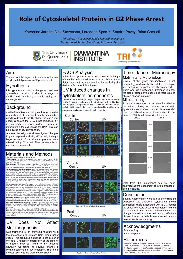
A2058 111nM 3000nM Role of Cytoskeletal Proteins in G2 Phase Arrest 37nM 333nM 1000nM MM576 Katherine Jordan, Alex Stevenson, Loredana Spoerri, Sandra Pavey, Brian Gabrielli The University of Queensland Diamantina Institute Translational Research Institute, Brisbane, Australia ^ Altered ^ FACS Analysis A FACS analysis was run to determine what length of time the cells should be exposed to UV for. It was determined that the optimum time for achieving the desired effect was 2:30 minutes. Aim The aim of this project is to determine the role of cytoskeletal proteins in G2 phase arrest. Hypothesis It is hypothesised that the change expression of cytoskeletal proteins is due to changes in motility, cell morphology, mitotic timing and melanogenesis. Time lapse Microscopy Motility and Morphology Several of the genes are implicated in cell morphology and motility. To test this, time lapse was performed on control and UV-B exposed. There was not a noticeable difference in either the size or shape of the cells, and there was no obvious change in motility. Mitotic timing A second movie was run to determine whether the mitotic timing was altered when actin dynamics were inhibited. Latrunculin A was also used to determine actin involvement in the process. 200nM will be used in the movie. Data from this experiment has not been analysed as the experiment is in the process of being repeated. A2058 UV induced changes in cytoskeletal components To determine the change in specific proteins after exposure to UV-B radiation cells were; fixed, stained with antibodies and imaged. Changes were found between UV and Control cells in cofilin, phalloidin, vimentin and paxillin, and between competent and defective cell lines in vimentin. MM576 Background Just before mitosis, a cell goes through a series of checkpoints to ensure it has the materials it needs to divide. In the G2 phase, there is a final check to ensure the DNA is not damaged. If it is, this leads to a pause, called G2 arrest, in mitosis while the cell repairs the DNA. This can be initiated by UV-B irradiation. A screen by Wigan et.al investigated changes in gene expression during G2 arrest, finding a large amount of cytoskeletal proteins were altered during G2 arrest. Their presence is not considered coincidental. UV Cofilin A2058 UV MM576 A2058 SKMel13 MM576 Control SKMel13 Materials and Methods Cell Lines - MM576, A2058, SKMeL13, HT144 FACS Analysis – All cell lines were plated into 5 dishes each. They were divided into 5 groups- control, 2 minutes UV- 24 hour examination, 2:00 UV/30 hours, 2:30/24 hours, 2:30/30 hours. Melanogenesis - The four cell lines were plated into 2 ten centimetre plates each. One plate from each cell line was exposed to UV radiation for 2:30, and twenty two hours later the cells moved to 1.5ml tubes and spun down in a centrifuge. Immunofluorescence/Fixation Method - Cells were plated onto coverslips and fixed using methanol, PFA 4% using Quench solution, PFA 3% using quench solution and finally PFA 3%. For methanol fixation, 4ml of -20°C Methanol was poured onto the cells. For all the PFA methods, 4ml of PFA (3% or 4%) was added. The cells were then incubated in PFA either on ice or at room temperature for 10, 15, 20, 30 or 40 mins. The PFA was removed and quench solution was then added and incubated for 10 mins, except for in the final 3% concentration where this step was skipped. After antibody staining (see below) the method which produced the clearest results was used. Immunofluorescence/Immunofluorescence - In the final staining, the cells were fixed with PFA3%, incubated for 15 minutes at room temperature and no quench solution was added. The cells were permeabilised with 0.1% Triton and incubated in blocking solution before the coverslips were placed into appropriate primary antibodies and incubated for 1 hour and 30 mins. They were then washed with 0.5% milk solution on a shaker for 3 x 10 mins before the coverslips were placed into secondary antibodies (mouse A-488 and rabbit A-555 and DAPI) and incubated for 35 minutes. The coverslips were then moved to slides and imaged. Time-lapse Microscopy 1 – 40,000 cells/well were plated into a 12-well plate, 2 wells for each cell line. One well from each cell line was exposed to UV radiation for 2:30 the next day. Eighteen hours later, the video was started and ran for 24 hours. Time-lapse Microscopy 2/Latrunculin Titration - Cell lines were plated 20,000 cells/well into 6 wells each in a 24 well plate. Latrunculin concentrations can be seen in results. The sixth well contained DMSO. Wells were imaged on inverted microscope. Time-lapse Microscopy 2/UV+Latrunculin - MM576 and A2058 were seeded into 4 wells each in a 12 well plate with 30,000 cells per well. Two days later two wells from each cell line were exposed to UV-B irradiation and one of these and a non-irradiated well had 1µM of latrunculin added. Time lapse microscopy began immediately and ran for 24 hours. Blue-DAPI, Green- α-Tubulin, Red- Cofilin Vimentin Control Conclusion Several experiments were run to determine the purpose of the change in cytoskeletal protein expression levels associated with a UV-induced G2 phase cell cycle arrest. It was determined that the change is not due to melanogenesis or a change in motility of the cell. It may affect the division time of the cells, however experiments to determine this are ongoing. Blue-DAPI, Green- α-Tubulin, Red- Vimentin Paxillin Control UV UV Does Not Affect Melanogenesis Melanogenesis is the polarising of granules in the melanocyte to protect DNA when under stress. This produces a change in the colour of the cells. Changes in expression of the proteins of interest may be linked to this process, however, we found no obvious change in melanin content after UV irradiation. This line of investigation was therefore not continued. Acknowledgments Sandrine Roy Fiona Simpson References Wigan M, Pinder A, Giles N, Pavey S, Burgess A, Wong S, Sturm RA, Gabrielli B (2012). A UVR-induced G2-phase checkpoint response to ssDNA gaps produced by replication fork bypass of unrepaired lesions is defective in melanoma. J Invest Dermatol.132(6):1681-8. Blue-DAPI, Green- α-Tubulin, Red- Paxillin