Download
1 / 54
790 likes | 1.57k Vues
Glycosaminoglycans. Dr Amena Rahim. Most mammalian cells are located in tissues where they are surrounded by a complex extracellular matrix (ECM) often referred to as “connective tissue.”. The ECM contains three major classes of biomolecules:

E N D
Glycosaminoglycans Dr Amena Rahim
Most mammalian cells are located in tissues where they are surrounded by a complex extracellular matrix (ECM) often referred to as “connective tissue.”
The ECM contains three major classes of biomolecules: (1) the structural proteins: collagen, elastin, and fibrillin; (2) certain specialized proteins such as fibrillin, fibronectin,and laminin; and (3) Proteoglycans
Functions: • Binding and packing of tissues (connective tissue proper) • Connect, anchor and support the body and its organs • Transport of metabolites between capillaries and tissues;
Defense against infection (via ground substance and cells) • Repair of injury (via cell proliferation and fiber formation) • Fat storage (as determined by age, sex, nutrition or disease)
The extracellular space in animal tissues is filled with a gel-like material, the extracellular matrix, also called ground substance, • which holds the cells of a tissue together and provides a porous pathway for the diffusion of nutrients and oxygen to individual cells.
Epithelial cells extra-cellularmatrix Underlying cells cells
The extracellular matrix is composed of an interlocking meshwork of heteropolysaccharides and fibrous proteins.
Heteropolysaccharides in the body are the glycosaminoglycans (GAGs). These molecules are long unbranched polysaccharides containing a repeating disaccharide unit.
GAGs are highly negatively charged molecules, with extended conformation that imparts high viscosity to the solution. • GAGs are located primarily on the surface of cells or in the extracellular matrix (ECM).
Along with the high viscosity of GAGs comes low compressibility, which makes these molecules ideal for a lubricating fluid in the joints.
At the same time, their rigidity provides structural integrity to cells and provides passageways between cells, allowing for cell migration.
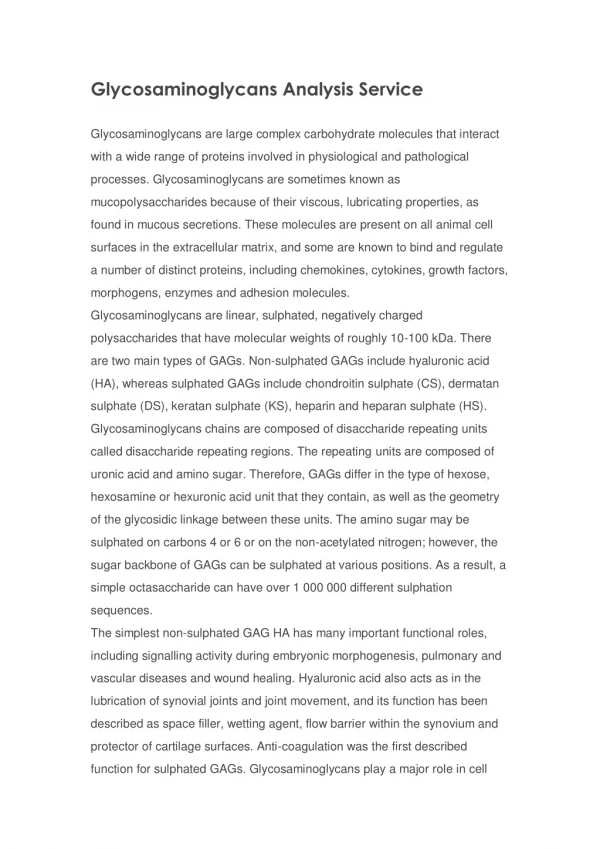
The disaccharide units contain either of two modified sugars, called amino sugarsN-acetylgalactosamine (GalNAc) or N-acetylglucosamine (GlcNAc), • and an acidic sugar uronic acid such as glucuronic acid or iduronic acid.
The amino group is usually acetylated. • This eliminates the positive charge.
In some glycosaminoglycans, one or more of the hydroxyls of the amino sugar is esterified with sulfate. • The combination of these sulfate groups and the carboxylate groups of the uronic acid residues gives the glycosaminoglycans a very high density of negative charge.
Keratan sulfate is an exception in which galactose is present, instead of an acidic sugar. • Hyaluronic acid does not contain sulfate.
Structure of Glycosaminoglycans • GAGs in the body are linked to core proteins ( except hyaluronic acid), forming proteoglycans (also called mucopolysaccharides).
The GAGs extend perpendicularly from the core in a brush-like structure. • E.g. in cartilage proteoglycan the GAGs present are chondriotin sulfate and keratan sulfate.
Linkage • The linkage of GAGs to the protein core involves a specific trisaccharide composed of two galactose residues and a xylose residue (Gal-Gal-Xyl-O-CH2-protein).
The trisaccharide linker is coupled to the protein core through an O-glycosidic bond to a Serine residue in the protein. • Some forms of keratan sulfates are linked to the protein core through an N-glycosidic bond.
The protein cores of proteoglycans are rich in Serine and Threonine residues, which allows multiple GAG attachments.
Proteoglycan Aggregates • Proteoglycan monomers associate with a molecule of hylauronic acid to form proteoglycan aggregates. • Association is not covalent but ionic between hyaluronic acid and the core protein. • Stabilized by link proteins
Classification of Glycosaminoglycans The classification is based on: OR the GAGs differ from each other: • Monomeric (acidic & amino sugar) composition • Degree & location of sulfation • Type of glycosidic linkages • Chain length of the disaccharides • Nature of the core protein • Their tissue distribution • Their biologic functions
The specific GAGs of physiological significance are: • Hyaluronic Acid • Dermatan Sulfate • Chondroitin Sulfate • Heparin • Heparan Sulfate • Keratan Sulfate
Characteristics of GAGs • Although each of these GAGs has a predominant disaccharide component , heterogeneity does exist in the sugars present in the make-up of any given class of GAG.
Hyaluronic acid • Hyaluronic acid is unique among the GAGs in that it does not contain any sulfate and is not found covalently attached to proteins as a proteoglycan. • It is, however, a component of non-covalently formed complexes with proteoglycans in the ECM.
Un sulfated • Only GAG present both in animals and bacteria.
Found in synovial fluid, • vitreous humor, • ECM of loose connective tissue • Umbilical cord • Cartilage
Specific function: 1. Hyaluronic acid is especially high in concentration in embryonic tissues and is thought to play an important role in permitting cell migration during morphogenesis and wound repair.
Association with major diseases: • Hyaluronic acid may be important in permitting tumor cells to migrate through the ECM. Tumor cells can induce fibroblasts to synthesize greatly increased amounts of this GAG, thereby perhaps facilitating their own spread
Chondroitin sulfate • most abundant GAG • Cartilage (bind collagen and hold the fibers strongly) • Tendons • ligaments • Heart valves
Heparan sulfate • Extracellular GAG • contains higher acetylated glucosamine than heparin • And less sulphated groups
found in the basement membrane of the kidney along with type IV collagen and laminin where it plays a major role in determining the charge selectiveness of glomerular filtration
are associated with the plasma membrane of cells, with their core proteins actually spanning that membrane. • In it they may act as receptors and may also participate in the mediation of cell growth and cell-cell communication.
Association with the disease: • Some tumor cells have less heparan sulfate at their surfaces, and this may play a role in the lack of adhesiveness that these cells display.
Heparin • It is an intracellular GAG. • Component of intracellular granules of mast cells lining the arteries of the lungs, liver and skin • more sulfated than heparan sulfate
Heparin is an important anticoagulant. It binds with factors IX and XI, but its most important interaction is with plasma antithrombin III.
Heparin can also bind specifically to lipoprotein lipase present in capillary walls, causing a release of this enzyme into the circulation.
Specific function: • Heparin and warfarin are widely used in the treatment of thrombotic and thromboembolic conditions, such as deep vein thrombosis and pulmonary embolus. • Heparin is administered first, because of its prompt onset of action, whereas warfarin takes several days to reach full effect.
Their effects are closely monitored by use of appropriate tests of coagulation because of the risk of producing hemorrhage.
Dermatan sulfate • Sclera- gives shape to the eye. • Binds LDL –plays a role in the development of atherosclerosis. • skin, blood vessels, heart valves
Keratan sulfate • cornea, • bone, • cartilage aggregated with chondroitin sulfates
Both keratan sulfate I and dermatan sulfate are present in the cornea. They lie between collagen fibrils and play a critical role in corneal transparency.
In various types of arthritis, proteoglycans may act as autoantigens, thus contributing to the pathologic features of these conditions. • The amount of chondroitin sulfate in cartilage diminishes with age.