Download
1 / 81
820 likes | 1.05k Vues
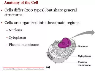
Anatomy of The Back I. Dr. Fadel Naim Orthopedic Surgeon Faculty of Medicine IUG. 1) Support weight Transmits weight to pelvis and lower limbs 2) Houses and protects spinal cord spinal nerves leave cord between vertebrae 3) Permits movements 4) Provides for muscle attachments

E N D
Anatomy of The Back I Dr. FadelNaim Orthopedic Surgeon Faculty of Medicine IUG
1) Support weight Transmits weight to pelvis and lower limbs 2) Houses and protects spinal cord spinal nerves leave cord between vertebrae 3) Permits movements 4) Provides for muscle attachments muscles of back muscles of head Neck upper extremity thorax Functions Of Vertebral Column
Formed from 33 bones in the adult Divided into five major regions Cervical vertebrae 7 vertebrae of the neck region Thoracic vertebrae 12 vertebrae of the thoracic region Lumbar vertebrae 5 vertebrae of the lower back Sacrum Inferior to lumbar vertebrae Articulates with coxal bones Coccyx Most inferior region of the vertebral column Regions and Normal Curvatures
Vertebral Parts lamina transverse process rib facet pedicle vertebral foramen body spinous process
Specific regions of the spine perform specific functions Types of movement that occur between vertebrae Flexion and extension Lateral flexion Rotation in the long axis Regions Vertebral Characteristics
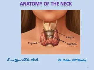
Seven cervical vertebrae (C1 – C7) smallest and lightest vertebrae C3 – C7 are typical cervical vertebrae Body is wider laterally Spinous processes are short and bifid (except C7) Vertebral foramen are large and triangular Transverse processes contain transverse foramina Superior articular facets face superoposteriorly Cervical Vertebrae
C1 is termed the atlas Lacks a body and spinous process Supports the skull Superior articular facets receive the occipital condyles Allows flexion and extension of neck Nodding the head “yes” The Atlas
Has a body and spinous process Dens (odontoid process) projects superiorly Formed from fusion of the body of the atlas with the axis Acts as a pivot for rotation of the atlas and skull Participates in rotating the head from side to side The Axis
Cervical Vertebrae bifid process Dens (odontoid process) transverse foramen Axis (C2) Atlas (C1)
Transitions to thoracic vertebrae has a long spinous process with a broad tubercle Not bifid Vertebra prominens (C7)
All articulate with ribs Have heart-shaped bodies from the superior view Each side of the body of T2 – T10 bears hemifacts for articulation with ribs T1 has a full facet for the first rib T11 – T12 only have a single facet Thoracic Vertebrae (T1 – T12)
Spinous processes are long and point inferiorly Vertebral foramen are circular Transverse processes articulate with tubercles of ribs Superior articular facets point posteriorly Inferior articular processes point anteriorly Allows rotation and prevents flexion and extension T10–T12 transition to lumbar vertebrae Thoracic Vertebrae
Bodies are thick and robust Transverse processes are thin and tapered Spinous processes are thick, blunt, and point posteriorly Vertebral foramina are triangular Superior and inferior articular facets directly medially Allows flexion and extension – rotation prevented Lumbar Vertebrae (L1 – L5)
Vertebral Column intervertebral disk intervertebral foamen
Thoracic & Lumbar Vertebrae lumbar vertebrae thoracic vertebrae
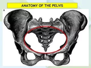
Shapes the posterior wall of pelvis Formed from 5 fused vertebrae Superior surface articulates with L5 Inferiorly articulates with coccyx Sacral promontory Where the first sacral vertebrae bulges into pelvic cavity Center of gravity is 1 cm posterior to sacral promontory Important in female Sacrum (S1 – S5)
Sacral foramina Ventral foramina Passage for ventral rami of sacral spinal nerves Dorsal foramina Passage for dorsal rami of sacral spinal nerves Sacrum
Characteristics : is curved, more in males than in females protects reproductive, urinary, and digestive organs Attaches the axial skeleton to pelvic girdle of appendicular skeleton Together ith iliac bone SI joint sacrum
The Sacrum • The adult sacrum: • consists of 5 fused sacral vertebrae • fuses between puberty and ages 25–30 • leaving transverse lines
Sacral hiatus: opening at the inferior end of the sacral canal formed by ridges of sacral cornua covered by connective tissues Sacral cornua: horn-shaped formed by laminae of the 5th sacral vertebra which do not meet at midline Sacral canal: replaces the vertebral canal
Median sacral crest: • fused spinous processes • Lateral sacral crest: • attach to muscles of lower back and hip • Auricular surface: • articulates with pelvic girdle (sacroiliac joint) • Sacral tuberosity: • attaches ligaments of the sacroiliac joint
4 Regions of the Sacrum • Base • the broad superior surface • Ala-wings • at either side of the base to attach muscles • Sacral promontory • at the center of the base • Apex • the narrow inferior portion articulates with the coccyx
Sacrum (5 fused vertebrae) sacral canal medial sacral crest sacral foramen sacral hiatus
Is the “tailbone” Formed from 3 – 5 fused vertebrae Offers only slight support to pelvic organs attaches ligaments and a constricting muscle of the anus Coccyx
The seventh cervical vertebra may possess a cervical rib The first lumbar vertebra, which may have a rib. Sacralization of 5th lumbar vertebra Lumberalization of 1st sacral vertebra A large extent of the posterior wall of the sacral canal may be absent The coccyx may have three or five vertebrae. The first coccygeal vertebra may be separate. IMPORTANT VARIATIONS IN THE VERTEBRAE
Joints between vertebrae in the back • A typical vertebra has a total of six joints with adjacent vertebrae: • four synovial joints (two above and two below) • two symphyses (one above and one below). • Each symphysis includes an intervertebral disc.
Movements of the spine are a function of the following features: • Size and compressibility of intervertebral discs • Tightness of joint capsules • Orientation of articular facets • Muscle and ligament function
Four distinct curvatures give vertebral column an S-shape Cervical and lumbar curvature concave posteriorly Thoracic and sacral curvatures convex posteriorly Curvatures increase the resilience of the spine Regions and Normal Curvatures
Curvatures of the vertebral column Primary curvatures thoracic and sacral curvatures An infant's spine is C-shaped at birth Secondary curvatures cervical and lumbar curvatures Develop when a baby begins to walk Redistributes weight of the upper body over the lower limbs The Axial Skeleton Throughout Life