It has been estimated that 90 percent of information used while driving is visual (Hills, 1980)
It has been estimated that 90 percent of information used while driving is visual (Hills, 1980) 20 % of U.S. adults mistakenly believe they do not need an eye exam unless they are having trouble seeing . This just isn’t true .*

It has been estimated that 90 percent of information used while driving is visual (Hills, 1980)
E N D
Presentation Transcript
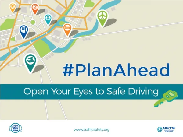
It has been estimated that 90 percent of information used whiledriving is visual (Hills, 1980) • 20% of U.S. adults mistakenly believe they do not need an eye exam unless they are having trouble seeing. This just isn’t true.* • Routine eye care is critical to healthy living, for numerous reasons! • * Americans’ Attitudes & Perceptions About Vision Care survey, 2012
How Do Eye Exams Improve My Driving? • Nearly all of the sensory input needed to drive comes from visual cues.* Your driving can be affected by: • distance vision – how quickly do you read signs and see hazards in front of you? • peripheral vision – how big is your blind spot? • near-vision focus – how legible is your dashboard? • night vision – can you readjust quickly from the glare? • color vision – can you see signs and lights clearly? • depth perception – how is your judgment when changing lanes and parking your car?
Many of these issues can go undetected without a regular full eye-exam. • Seeing well helps you drive well!
How often do you need an eye exam? • The American Optometric Association recommends: • 18-60 years old with no risk factors: an eye exam every two years • 18-60 years old with family history of eye disease, diabetes, high blood pressure, taking certain medications or contact lens use: more frequent exams may be necessary -- always follow the recommendation of your eye doctor • 61+ years old: an annual, comprehensive eye exam that includes testing for cataracts, glaucoma, macular degeneration, diabetic retinopathy and other conditions commonly associated with aging
Vision and Safety Eyewear Guidefor U.S. Army Civilian and Military Job Series
Visual acuity Snellen chart Acuity at Near&Distance
Near visual acuity • Definition • The ability to see clearly at a normal reading distance • Method of measuring • Jaeger card-reduced snellen chart • Causes of decreased near V.A in the presence of normal distance V.A • Presbyopia,uncorrectedhyperopia,centerally located cataracts and drug side effects
Visual field Confrontation test primetry
Visual field • Normal extension • 70 degrees Inferiorly, 60 degrees superiorly,95 degrees temporally and 60 degrees nasally. Total horizontal visual field extent to 190 degrees • Factors that influence the visual field • Size, distance, background illumination, colour, Kind of job, Bi or monocular vision, age, use of Personal protective device ( safety spectacle)
Normally, there are three kinds of cones (each one sensitive to a specific range of wavelengths): "red" cones (64%) "green" cones (32%) "blue" cones (2%) Color blindness The normal human retina contains two kinds of light sensitive cells: the rod cells (active only in low light) and the cone cells (active in normal daylight and responsible for color perception). The different kinds of inherited color blindness result from partial or complete loss of function of one or more of the different cone systems.
Different Types of Color Blindness • Monochromacy: occurs when two or all three of the cone pigments are missing and color and lightness vision is reduced to one dimension. • Total color blindness • Dichromacy: occurs when only one of the cone pigments is missing and color is reduced to two dimensions. • Partial color blindness red-green blue-yellow
When a person is tested using the Ishihara test, a score of ten correct answers out of eleven is considered to be within the normal range. A score of seven or less out of eleven is abnormal, and the person would be considered to have this deficiency of the ability to correctly see colors. If the score is either eight or nine out of a possible eleven correct answers, further testing will be needed to find if the person is truly exhibiting red green deficient tendencies.
Most cases of congenital color vision deficiency are characterized by a red-green deficiency which may be of two types; first, a protan type which may be complete (protanopia) or partial (protanomalia), and, secondly, a deutan type which may be complete (deuteranopia) or partial (duteranomalia).
Photoreceptor Anatomy • Example: if you stimulate all 3 types of cones about equally the result is white or no color.
Colour vision test Ishihara plates
October 2001 Taylor, JF (1987). Vision and Driving. Ophthal. Physiol Opt. Vol 7, No. 2, pp 187-189. Road Traffic Act (1988). London HMSO. Highway Code London HMSO Colour vision Impaired colour vision is not a bar to driving.
Depth perception (Stereopsis) • The ability to precieve depth or relative distance Some jobs that need stereoscopic vision • Furk-lift truck operator , crane driver, pilot ,… Factors that influence depth perception • Uncorrected refractive errores, amblyopia , squint low level of illumination , anisometropia , age , Advantages of binocular vision over monocular • The presence of stereopsis , improved visual acuity , an enlarged Field of peripheral vision , slightly brighter of object
Clinical tests for stereopsis Contour stereotest • Titmusstereotest Random dot stereotest • Frisby test • Random Dot-E test
Clinical Contrast Sensitivity Tests • Pelli-Robson chart • Regan low-contrast • Vistech chart • Melbourne edge test
Distance vision disorders • In most states in the United States (42 States or 84 percent), an unrestricted private driver’s license requires visual acuity of 20/40 or better (corrected or uncorrected) in one eye alone. In other countries (e.g., Europe, Asia, Africa, the Middle East, • Australia), the most prevalent acuity standard is 20/40 combined, or both eyes tested together (Keeney, 1993).
Cataract • . After adjustment for driving exposure, results revealed that drivers with cataracts were 2.5 times more likely to have a history of at-fault crashes in the previous five years compared to those without cataract
colour vision • Despite the reported difficulties with colour vision discrimination while driving, it is unlikely that colour vision impairments, in general, represent a driving hazard, particularly now that the position of traffic lights has been generally standardized (Canadian Medical Association, 1999). Nevertheless, it seems prudent to caution drivers regarding potentially hazardous situations (e.g., traffic lights, brake lights, parked cars).
contrast sensitivity • In general, the research that is available suggests that impairments in contrast sensitivity are associated with higher rates of crashes. Results from Owsley (2000) suggest that measures of contrast sensitivity may be a more sensitive predictor of crash risk than current measures of visual acuity. Although more research is needed, it may be prudent to include measures of contrast sensitivity in assessments of older drivers.
Diabetic Retinopathy • Treatment of proliferative diabetic retinopathy by pan retinal photocoagulation (laser ablation of the retina) can cause reduced visual field and jeopardies the right to drive. DVLA will investigate fields when laser treatment has been carried out on both eyes.
glaucoma • the frequency of visual field loss in individuals with glaucoma was 10 times greater than for the general population examined. Results of the study also indicated that individuals with a family history of glaucoma had a higher incidence (5.6 percent) of visual field loss compared to the general population studied(age- and sex-matched controls).
glaucoma • Important to this review was the finding that individuals with visual field loss in both eyes had double the crash and conviction rates (per person per 160,000 km) as an age- and sex-matched control group with normal visual fields. The crash and conviction rates of those with monocular visual field loss were not significantly
monocular • The authors concluded that despite the reductions in selective visual functions, monocular drivers are not significantly worse than binocular drivers in the safety of most day-to-day driving functions. Measures of crash rates were not included in the study.
ARMD • . Finally, results of the study indicated that the age- related macular degeneration group compensated for their impairments in four ways: not driving in unfamiliar areas, traveling at slower speeds, self- restricting their night time driving, and taking fewer risks while driving (e.g., not changing lanes)