Download
1 / 1
10 likes | 118 Vues
Explore the interaction between RhoGAP proteins and their targets, Rho, Cdc42, and Rac, utilizing a molecular graphics program. Investigate key structural motifs crucial in this interaction and identify potential RhoGAP targets among structural homologs.

E N D
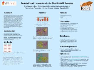
Protein-Protein Interaction in the Rho-RhoGAP Complex Eno Akpovwa, Paul Craig, Herbert Bernstein, Rochester Institute of Technology, Rochester, NY and Dowling College, Oakdale, NY. Abstract Results Results The RhoGAP (Rho GTPase Activating Protein) family of proteins regulates G-protein signal transduction cascades by increasing the slow intrinsic GTP to GDP hydrolysis by Rho. The Rho family of small G proteins tranduces signals from plasma-membrane receptors and controls cell adhesion, motility and shape by actin cytoskeleton formation. This process occurs through transient protein-protein interactions between RhoGAP and its targets, Rho, Cdc42 and Rac. Rho and its homologs interact with RhoGAP by packing together with the RhoGAP protein, so that their switch I and II regions slot into a shallow pocket on RhoGAP, making polar and non-polar interactions with RhoGAP. Using a molecular graphics program (PyMol) with a plug-in designed at RIT (ProMol); the small structural motifs involved in the Rho-RhoGAP interaction have been explored. Two RhoGAP motifs were created – one was based on conserved residues involved in protein-protein interactions and the second was based on five residues that promote GTP hydrolysis. These motifs were then tested using a series of RhoGAP homologs. The results demonstrated that the Motif Maker tool in ProMOL is able to discriminate between RhoGAP homologs that actively target Rho and those homologs that share fold similarity with RhoGAP, but are not able to interact with and activate Rho. • 300 Random PDBs (protein structures from the protein data base). • Of the 300 random PDBs, only 3% were structural homologs. • None was a RhoGAP Target. Discussion • Homologs that are not RhoGAP targets, but structural homologs show some residues from the PPI motif. • Also, there are some homologs which are not structural homologs and RhoGAP targets, but show residues from the PPI motif. • Some of the homologs showing residues from the PPI motif were equivalent to residues in the Ras protein. • There were some homologs with a couple of the residues from the PPI motif that did not align with the residues in the PPI motif. • Non-structural homologs have residues from the PPI motif. • This suggests sequence homology, but might have different functions. • Structural homologs but not RhoGAP target show residues from PPI motif. • This suggests there might be more RhoGAP targets that have not being explored yet. • Homologs with the residues in the sequence that do not align might have a different function. Figure 2: Data from comparing 324 homologs against the RhoGAP-Rho motif Introduction The purpose of this research was to determine if a molecular graphics program, such as PyMol can help determine protein-protein interactions through their binding domains. Hypothesis: Using a molecular graphic program (PyMol) and a plug-in ProMOL( a motif maker), RhoGAP targets and other possible homologs which can be possible RhoGAP targets can be identified. Conclusion Figure 3: Alignment of Rho (yellow) and a structural homolog of Rho (magenta) that is also a RhoGAP target according to the literature and ProMOL (residues align on each other). • Of all homologs studied, 55% were structural homologs of Rho. • Of the 55%, only 10% were RhoGAP targets according to the literature. • However, ProMOL identified 26% of the homologs studied as RhoGAP targets. • Structural homologs which are not RhoGAP targets, but show residues can be tested in the lab to determine if they are potential RhoGAP targets. Methods Acknowledgements This work was supported by the RIT College of Science and funding from NIGMS 1 R15 AG16192-01 and NIGMS 3 R15 GM078077-01S1. I would also like to thank Joyce Murphy for her help, encouragement and support. Figure 4: Alignment of Rho (yellow) and a structural homolog that is not a RhoGAP target (magenta, residues do not align). References Figure 1: Residues important for RhoGTPase Activating Protein to bind to its target, Rho. Barker, J. et al. Catalytic Site Atlas, http://www.ebi.ac.uk/thornton-srv/databases/CSA/. Accessed 7 Aug. 2009 Basic Local Alignment Search Tool. http:/// blast.ncbi.nlm.nih.gov/Blast.cgi (accessed 5 Aug 2009). RCSB Protein Data Bank. http://www.rcsb.org/pdb/home/home.do (Accessed 5 Aug. 2009). Letunic, I., Doerks, T., Bork, P. SMART 6: recent updates and new developments. Nucleic Acids Research 37: D229-232 (2008). Barrett, T. et al. "The structure of the GTPase-activating domain from p50rhoGAP.” Nature 385: 458-461 (1997). Rittinger, K. et al. "Crystal Structure of a small G protein in complex with the GTPase-activating protein rhoGAP." Nature 388: 693-697 (1997). Rittinger K. et al. "Structure at 1.65 A of RhoA and its GTPase-activating protein in complex with a transition-state analogue.” Nature 389: 758-762 (1997). Using the SMART database, Protein Data Bank and Catalytic Site Atlas, a motif was made by first looking for PDB entries that have either the RhoGAP or Rho protein in them. Then, these files were opened using PyMOL. With the ProMOL plug-in, the important amino acid residues needed in the RhoGAP and Rho protein were used to create a motif. Subsequently, PDB entries that may be Rho or RhoGAP homologs can be identified using these motifs. Figure 5: Alignment of Rho (yellow) and a related structure that is neither a structural homolog or a RhoGAP target (backbone and residues don’t align).