Download
1 / 17
180 likes | 288 Vues
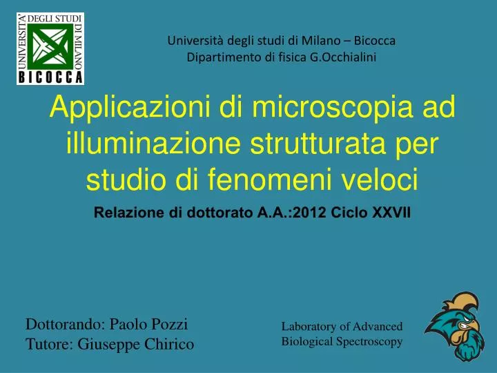
Università degli studi di Milano – Bicocca Dipartimento di fisica G.Occhialini. Applicazioni di microscopia ad illuminazione strutturata per studio di fenomeni veloci. Relazione di dottorato A.A.:2012 Ciclo XXVII. Dottorando : Paolo Pozzi Tutore : Giuseppe Chirico.

E N D
Universitàdeglistudi di Milano – Bicocca Dipartimento di fisicaG.Occhialini Applicazioni di microscopia ad illuminazionestrutturata per studio di fenomeniveloci Relazionedi dottoratoA.A.:2012 Ciclo XXVII Dottorando: Paolo Pozzi Tutore: Giuseppe Chirico Laboratory of Advanced Biological Spectroscopy
Summary • Fluorescence • “Standard” Microscopy • Structured Illumination • Neural Network Analysis • Calcium Imaging
Fluorescence S2 fluorophore S1 Linear phenomenon
Epifluorescence Microscope CCD Emission filter Lamp Dichroic mirror Excitation filter Microscope Field of View Objective Sample
Two Photon Fluorescence S2 fluorophore S1 Non - Linear phenomenon
Two Photon Microscope Phototube Emission filter Galvanometric mirrors Dichroic mirror Laser Source Microscope Field of View Objective Sample
Scanning Imaging Very slow!!! 10-15 fps max I t
Phase Shaping Standard microscope: FFT Structured Illumination: FFT
Structured Illumination Microscope CCD Emission filter Laser Source Dichroic mirror Microscope Field of View SLM Objective Sample
What do we Watch? Millisecond scale fluorescence variations in multiple locations: Blood flow cross-correlation Neural network monitoring
Neurons Dendrites Ionic Channels Axon
Action Potential Potassium Channels Open Potential Sodium Channels Open Resting potential Time s
Voltage Sensitive Dyes • Membrane Marker • Extremely sensible to Stark Effect • Two photon fluorescence change S2 S1
The experiment Mossy Fiber (Input) Granule Cells (Elaboration) Purkinje Cell (Output)
Data Acquisition WIP 5