Download
1 / 66
660 likes | 699 Vues
Explore the essential functions and different tissues of the skeletal system, such as bone and cartilage, along with the structural components like compact and spongy bone. Delve into the composition of bone, cellular components, and bone structure.

E N D
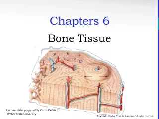
Chapters 6 Bone Tissue Lecture slides prepared by Curtis DeFriez, Weber State University
Introduction • The skeletal system has 6 important functions: • Provide support by acting as a structural framework and a point of attachment for tendons and ligaments • Protect the internal organs (brain, chest, etc.) • Assist body movements (in conjunction with muscles) • Store and release salts of calcium and phosphorus • Participate in blood cell production (hematopoiesis) • Store triglycerides in adipose cells of yellow marrow
Tissues of the Skeletal System • Bone is a dynamic tissue – it is always remodeling (building up and breaking down). • Like all organ systems (and as part of the even larger musculoskeletal organ system), the skeletal system is made of several different tissues. • The two major tissues are bone (osseous tissue) and cartilage.
Tissues of the Skeletal System • Bone is a highly vascularized C.T. with a hard, mineralized extracellular matrix. It is found in the body in two different arrangements: • Compact bone – most of the bone in this graphic is compact bone. • Spongy bone is seen as the less organized tissue along the left margin (with the spicules).
Tissues of the Skeletal System • Compact bone is good at providing protection and support. • It forms the diaphysis of long bones, and the external layer of all bones. • Spongy bone is lightweight and provides tissue support . • It forms much of the epiphysis and the internal cavity of long bones. Compact bone Spongy bone
Tissues of the Skeletal System • Cartilage is a poorly vascularized C.T. with a matrix composed of chondroitin sulfate and various fibers. • Fiber types distinguish hyaline cartilage from fibrocartilage or elastic cartilage. Hyaline cartilage
Tissues of the Skeletal System • Articular cartilage is the thin layer of hyaline cartilage covering the epiphysis of long bones. • Articular cartilage is found where the bone forms an articular (joint) surface - where one bone moves against another bone. Hyaline cartilage is the articular cartilage of this long bone
Tissues of the Skeletal System • The periosteum is a tough sheath of dense, irregular connective tissue on the outside of the bone. • It contains osteoblasts that help the bone grow in thickness, but not in length. • It also assists with fracture repair and serves as an attachment point for tendons and ligaments.
Structure of Bone • The medullary cavity is a space within the diaphysis of long bones that contains fatty yellow bone marrow in adults. • The endosteum is a membrane that lines the medullary cavity . • The endosteum is composed of osteoclasts, osteoblasts, and connective tissue.
Tissues of the Skeletal System • The perichondrium is a dense irregular connective tissue membrane that surrounds cartilage. • Chondrocytes are cells that form cartilage. • As we will soon see, many of the major bones are formed from cartilage (the remainder do not go through a cartilaginous stage.) Perichondrium Periosteum
Tissues of the Skeletal System • The various cells in osseous tissues are shown in the bottom graphic:
Tissues of the Skeletal System • Osteoblasts are bone building cells: They synthesize and secrete collagen fibers and other organic components. • Osteocytes are mature osteoblasts (maintenance). • Osteoclasts are large bone breakdown cells. • As white blood cells, osteoclasts migrated from the bone marrow to become “fixed macrophages” in the substance of the bone.
Tissues of the Skeletal System • Besides bone and cartilage, the skeletal system contains other important tissues: • Epithelium (endothelium) form the capillary walls • Nerves (the periosteum is especially tender) • Red marrow – hematopoiesis • Yellow marrow – fat storage
Chemical Constituents of Bone • Bone is 25% water, 25% organic proteins, 50% mineral salts (hydroxyapatite crystals). • Organic constituents • Collagen fibers provide flexibility and tensile strength. • Inorganic hydroxyapatite crystals (mineral salts) • Calcium Phosphate (Ca3PO4)2 • Calcium Carbonate (CaCO3 – marble) • Other trace elements: magnesium, fluoride, sulfate
Bone Structure • The humerus in the arm is a typical long bone.
Bone Structure • The diaphysis is the shaft or body of a long bone. • The epiphyses form the distal and proximal ends of a long bone. • The metaphyses are the areas where the epiphyses and diaphysis join.
Bone Structure • In adolescents, through the end of active growth, the epiphysis of the long bones contains hyaline cartilage and forms an “epiphyseal growth plate”. • The growth plate is always actively dividing and causing the bone to elongate from each end.
Bone Structure • In adults, the epiphyseal cartilage is no longer present and elongation of bones has stopped. • The epiphyseal growth plate becomes an “epiphyseal line”, as growing cartilage is replaced by calcified bone. • The epiphyseal line is visible externally and on X-rays.
Histology of Bone Tissue • Compact Bone contains units called osteons or Haversian systems formed from concentric lamellae (rings of calcified matrix). • Interstitial lamellae between osteons are left over fragments of older osteons.
Histology of Bone Tissue • Outer circumferential lamellae encircle the bone beneath the periosteum. • Inner circumferential lamellae encircle the medullary cavity.
Histology of Bone Tissue • Lacunae are small spaces between the lamellae which house osteocytes. • Canaliculi are small channels filled with extracellular fluid connecting the lacunae.
Histology of Bone Tissue • Blood and lymphatic vessels are found in the osteon’s Central canal. • Perforating (Volkmann’s) canals allow transit of these vessels to the outer cortex of the bone.
Histology of Bone Tissue • Spongy bone lacks osteons. Instead, lamellae are arranged in a lattice of thin columns called trabeculae. • Trabeculae of spongy bone support and protect the red bone marrow and are oriented along lines of stress (helps bones resist stresses without breaking). • Hematopoiesis (blood cell production) occurs in spongy bone.
Histology of Bone Tissue • Within each trabecula of spongy bone are lacunae . • As in compact bone, lacunae contain osteocytes that nourish the mature bone tissue from the blood circulating through the trabeculae.
Histology of Bone Tissue • The interior of long bones is made up primarily of spongy bone. The use of spongy bone lessens overall bone weight.
Blood and Nerve Supply of Bone • Bone is richly supplied with blood; Periosteal arteries and veins supply the periosteum and compact bone. • Nerves accompany the blood vessels (this is often the case.) • The periosteum is rich in sensory nerves sensitive to tearing or tension (as anyone who has bruised their shin will tell you!)
Bone Formation • Ossification or osteogenesis is the process of forming new bone. Bone formation occurs in four situations: • Formation of bone in an embryo • Growth of bones until adulthood • Remodeling of bone • Repair of fractures
Bone Formation • Osteogenesis occurs by two different methods, beginning about the 6th week of embryonic development. • Intra-membranous ossification produces spongy bone. • This bone may subsequently be remodeled to form compact bone. • Endochondral ossification is a process whereby cartilage is replaced by bone. • Forms both compact and spongy bone.
Bone Formation • Intra-membranous ossification is the simpler of the two methods. • It is used in forming the flat bones of the skull, mandible, and clavicle. • Bone forms from mesenchymal cells that develop within a membrane – without going through a cartilage stage (recall that mesenchyme is the tissue from which almost all other C.T. develop.)
Bone Formation • Endochondral ossification is the method used in the formation of most bones, especially long bones. • It involves replacement of cartilage by bone. • There are one primary and two secondary centers of growth. Which means 2 different types.
Bone Formation • Ossification contributing to bone length is usually complete by 18-21 years of age. • Bones can still continue to thicken and are capable of repair even after the epiphyseal growth plates have closed.
Bone Formation • Human growth hormone is one of the body’s many anabolic hormones. Among other things, its secretion will stimulate bone growth, muscle growth, loss of fat, and increased glucose output in the liver. • The use of growth hormone has been increasing in popularity among athletes due to the numerous “benefits” associated with its use; side effects are often not thought of when young athletes use these drugs.
Bone FormationInteractions Animation • Bone Formation You must be connected to the internet to run this animation
Bone Growth and Remodeling • A balance must exist between the actions of osteoclasts and osteoblasts. • If too much new tissue is formed, the bones become abnormally thick and heavy (acromegaly). • Excessive loss of calcium weakens the bones, as occurs in osteoporosis. • They may also become too “soft”, as seen in the bone diseases rickets and osteomalacia.
Bone Growth and RemodelingInteractions Animation • Bone Remodeling You must be connected to the internet to run this animation
Bone Growth and Remodeling Normal bone metabolism depends on several factors: • Minerals are an essential component. • Large amounts of calcium and phosphorus and smaller amounts of magnesium, fluoride, and manganese are required for bone growth and remodeling.
Bone Growth and Remodeling • Vitamins are also necessary for normal bone metabolism: • Vitamin A stimulates activity of osteoblasts. • Vitamin C is needed for synthesis of collagen. • Vitamin D is essential to healthy bones because it promotes the absorption of calcium from foods in the gastrointestinal tract into the blood. • Vitamins K and B12 are needed for synthesis of bone proteins.
Bone Growth and Remodeling • Hormones are key contributors to normal bone metabolism. • During childhood, the hormones most important to bone growth are human growth hormone (hGH) and growth factors called IGFs (produced by the liver). Both stimulate osteoblasts, promote cell division at the epiphyseal plate, and enhance protein synthesis. • Thyroid hormones and insulin also promote bone growth by stimulating osteoblasts and protein synthesis.