
Fluorescent Imaging of MS Cells with Phalloidin and pMLC Staining
This study presents a detailed analysis of MS cells stained with Phalloidin (Red), highlighting cytoskeletal structures, and phosphorylated myosin light chain (pMLC) (Green) through fluorescence microscopy. The images capture the intricate organization and interaction of cell components under varying conditions, represented in the merged images for better visualization. Different cell lines (dlx1G, dlx1E, dlx1D, and dlx1A) are included, showcasing the dynamic nature of cytoskeletal regulation by pMLC phosphorylation.

Fluorescent Imaging of MS Cells with Phalloidin and pMLC Staining
E N D
Presentation Transcript
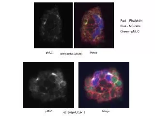
Red – Phalloidin Blue - MS cells Green - pMLC pMLC Merge 021908pMLCdlx1G pMLC Merge 021908pMLCdlx1E
Red – Phalloidin Blue - MS cells Green - pMLC pMLC Merge 021908pMLCdlx1D
Red – Phalloidin Blue - MS cells Green - pMLC pMLC Merge 021908pMLCdlx1A