Chapter 30
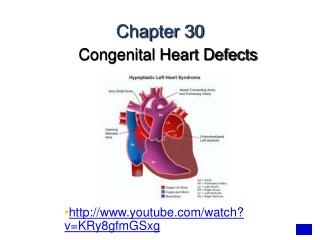
Chapter 30. Congenital Heart Defects. http://www.youtube.com/watch?v=KRy8gfmGSxg. Cardiac Defects. Patent Ductus Arteriosus Atrial Septal Defect Ventricular Septal Defect Tetralogy of Fallot Transposition of the Great Arteries Coarctation of the Aorta Anomalous Venous Return

Chapter 30
E N D
Presentation Transcript
Chapter 30 Congenital Heart Defects • http://www.youtube.com/watch?v=KRy8gfmGSxg
Cardiac Defects • Patent Ductus Arteriosus • Atrial Septal Defect • Ventricular Septal Defect • Tetralogy of Fallot • Transposition of the Great Arteries • Coarctation of the Aorta • Anomalous Venous Return • Truncus Arteriosus • Hypoplastic Left-Heart Syndrome
Heart • Congenital heart disease (CHD) occurs in 1/125 live births. • Neonates may present with a variety of non-specific findings, including: - tachypnea - cyanosis - pallor - lethargy - FTT - sweating with feeds • More specific findings include: - pathological murmurs - hypertension - abnormal pulses - syncope
Neonatal cardiac physiology • The transformation from fetal to neonatal circulation involves two major changes: • A marked increase in systemic resistance. • caused by loss of the low-resistance placenta. 2. A marked decrease in pulmonary resistance. • caused by pulmonary artery dilation with the neonate’s first breaths.
Fetal Circulation • No circulation to lungs • Foramen ovale • Ductus arteriosum • Circulation must go to placenta • Umbilical aa., vv.
Fetal cardiac physiology Fetal circulation: • Blood flows from the placenta IVC RA through the PFO LA LV • ascending aorta • brain • returns via the SVC
Fetal cardiac physiology Fetal circulation: • From the SVC RA RV • pulm aa • through the PDA • descending aorta • lower extremities and placenta
Fetal cardiac physiology Fetal circulation: • Only a very small amount of blood is directed through the right and left pulmonary aa’s to the lungs.
Neonatal cardiac physiology Neonate circulation: • The transformation to neonatal circulation occurs with the first few breaths. • The two remaining remnants of the fetal circulation are a patent foramen ovale... • and ductus arteriosus.
Congenital Heart Disease • Neonates with CHD often rely on a patent ductus arteriosus and/or foramen ovale to sustain life. • Unfortunately for these neonates, both of these passages begins to close following birth. • The ductus normally closes by 72hrs. • The foramen ovale normally closes by 3 months. • http://www.youtube.com/watch?v=FG-CNV501bc
CHD • That being said, in the presence of hypoxia or acidosis (generally present in ductus-dependent lesions), the ductus may remain open for a longer period of time. • As a result, these patients often present to the ED during the first 1-3 weeks of life. • i.e. as the ductus begins to close.
Classifying CHD • There are many different classification systems for CHD. • None are particularly good. • I will be discussing the Pink/Blue/Grey-Baby system: • Pink Baby – Left to right shunt • Blue Baby – Right to left shunt • Grey Baby – LV outflow tract obstruction
Pink Baby (L R shunt) • L R shunts cause CHF and pulmonary hypertension. • This leads to RV enlargement, RV failure, and cor pulmonale. • These babies present with CHF and respiratory distress. • They are not typically cyanotic. • http://www.youtube.com/watch?v=46tmI2_RVuE&list=PLA81DD78BDE77FBFC
Pink Baby (L R shunt) • These lesions include (among others) ASD’s, VSD’s, and persistently patent ductus arteriosus. • VSD • ASD
Pink Baby (L R shunt) • Persistently patent ductus arteriosus
Pink Baby (L R shunt) • Diagnosing L R shunts depends on: 1. Examination findings: • Non-cyanotic infant in resp distress. • Crackles, widely-fixed second heart sound, elevated JVP, cor pulmonale. 2. CXR: • Increased pulmonary vasculature (suggestive of CHF). • RA and/or RV enlargement.
Pink Baby (L R shunt) • Initial management should be directed at reducing the pulm edema. • Adminster Lasix 1mg/kg IV. • Peds Cardiology/ PICU should be consulted urgently regarding use of: • Morphine • Nitrates • Digoxin • Inotropes
Ductus Arteriosus • Fetal Circulation Component • Connects Pulmonary Artery to Aorta • Shunts blood away from lungs • Maintained patent by presence of prostaglandins • Closure secondary to: • Increase in PaO2_ • Decrease in level of prostaglandins
Patent Ductus Arteriosus • 5-10% of all births (1 of 2000 live births) • 80% of premature babies • 2-3 times more common in females than males. • 5th or 6th most common congenital cardiac defect. • Often associated with other defects. • May be desirable with some defects. • Morbidity/Mortality related to degree of blood flow through PDA.
Pathophysiology - PDA • With a drop in pulmonary arterial pressure (reduction in hypoxic pulmonary vascular constriction), blood will flow through PDA. • LEFT TO RIGHT SHUNT • Increased pulmonary blood flow may lead to pulmonary edema. • Reduced blood flow to all postductal organs • NEC • If pulmonary artery pressure rises above Aortic pressure, blood will move in the other direction. • RIGHT TO LEFT SHUNT
Diagnosis - PDA • Loud grade I to grade III systolic murmur at left sternal border. • Washing machine • Echocardiography
Treatment - PDA • Restrict fluids. • Diuretics • Prostaglandin Inhibitors - Indomethacin • Surgical closure (ligation).
Atrial Septal Defect • 6-10% of all births (1 of 1500 live births) • 2 times more common in females than males. • Types: • Ostium Secundum (at or about the Foramen Ovale) • Sinus Venous • In 1950 most children with ASD did not reach the first grade. Today, first year surgery facilitates normal growth and development.
ASD: Pathophysiology and Diagnosis • Pathophysiology • Left to Right Shunt • Inefficient recirculation of good blood through pulmonary arteries. • May not manifest symptoms and may be found later in life. • If defect is significant, may cause problems later in life due to inefficiencies. • Diagnosis • Murmur • Echocardiography
Treatment - ASD • Surgical closure. • Non-Surgical closure via cardiac catheterization.
Ventricular Septal Defect • 1% of all births (2 to 4 of 1000 live births) • Vast majority the hole is small. • In 1950, fatal. Today almost all VSD can be closed successfully, even in small babies. Lillehei was the first person in history to correct both ASD and VSD on 8/31/54.
VSD: Pathophysiology & Diagnosis • Pathophysiology • May be isolated or associated with other congenital cardiac defects. • With normal PVR: • LEFT TO RIGHT SHUNT • With elevated PVR (RDS): • RIGHT TO LEFT SHUNT • Diagnosis • Echocardiography
Treatment - VSD • Nothing if VSD is small. • With CHF or Failure to Thrive: Surgical closure.
Blue Baby (R L shunt) • R L shunts cause hypoxia and central cyanosis. • Neither hypoxia or cyanosis tend to improve with 100% oxygen. • R L lesions include (among others): • Tetralogy of Fallot (TOF) • Transposition of the Great Arteries (TGA)
Blue Baby (R L shunt) • Hypoxia and cyanosis (unresponsive to oxygen) in the neonatal period suggests a ductus-dependent lesion. • Treatment is a prostaglandin-E1 (PGE1) infusion. • Dosing discussed momentarily • This should obviously be accompanied by urgent Peds Cardiology and PICU consultation.
Tetralogy of Fallot • Characterized by: • Pulmonary aa OTO • RV hypertrophy • VSD • Over-riding aorta • With severe pulmonary OTO... • * • * • * • bloodflow to the lungs may be highly ductus-dependent. • *
Tetralogy of Fallot • The classic CXR finding in TOF is the boot-shaped heart. • Pulmonary vasculature is typically decreased.
Tetralogy of Fallot • 1% of neonates. • Most common of the cyanotic cardiac diseases. • Mortality increases with age (1 year-old has a 25% mortality, 40 year-old has 95%). • In 1950, fatal. Today, less than 5% mortality with children operated on in infancy, leading normal lives.Four Defects • Pulmonary Artery Stenosis (determinant factor related to severity) • VSD (usually large) • Overriding Aorta • RV hypertrophy
Tetralogy of Fallot: Diagnosis and Treatment • Tet Spells (Blue spells) • CXR: Boot-shaped Heart • Diagnosed with echocardiography. • Surgical correction. • Reparative or Palliative (Blalock-Taussig)
Blalock-Taussig • Something the Lord Made. • Vivien Thomas
Transposition of the Great Arteries • TGA is one of the most common cyanotic lesion presenting in the first week of life. • Anatomically: • RV aorta • LV pulmonary aa • To be compatible with life, mixing of the two circulations must occur via an ASD, VSD, or PDA. • http://www.youtube.com/watch?v=O83cYwKOKtI&list=PLA81DD78BDE77FBFC
Transposition of the Great Arteries • The CXR findings in TGA are typically less dramatic than in TOF. • Pulmonary vasculature is typically increased. • http://www.youtube.com/watch?v=b-TkE_wygT4&list=PLA81DD78BDE77FBFC&index=1
Complete Transposition of the Great Arteries • Second most common form (5-7%) of congenital cardiac anomalies. • Aorta arises from RV and Pulmonary Arteries from LV. • Without an abnormality, life would not be possible. • ASD • VSD (30-40%) • PDA