Download
1 / 20
220 likes | 439 Vues
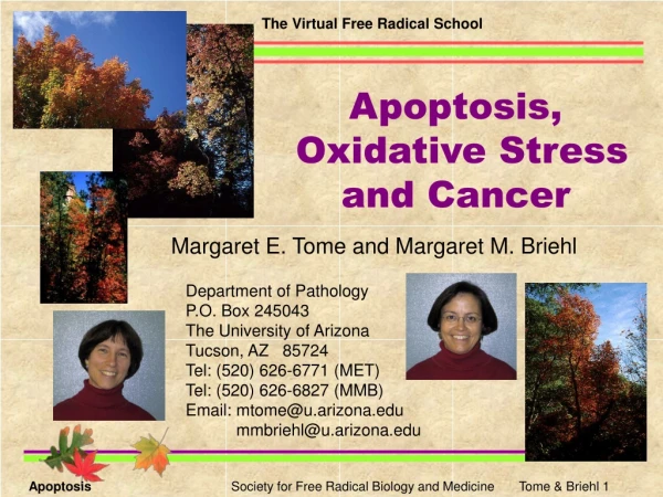
The Virtual Free Radical School. Apoptosis, Oxidative Stress and Cancer. Margaret E. Tome and Margaret M. Briehl. Department of Pathology P.O. Box 245043 The University of Arizona Tucson, AZ 85724 Tel: (520) 626-6771 (MET) Tel: (520) 626-6827 (MMB) Email: mtome@u.arizona.edu

E N D
The Virtual Free Radical School Apoptosis, Oxidative Stress and Cancer Margaret E. Tome and Margaret M. Briehl Department of Pathology P.O. Box 245043 The University of Arizona Tucson, AZ 85724 Tel: (520) 626-6771 (MET) Tel: (520) 626-6827 (MMB) Email: mtome@u.arizona.edu mmbriehl@u.arizona.edu Apoptosis Oxygen Society Education Program Tome & Briehl 1
Apoptosis - (Gr. "falling") a process seen in multicellular organisms, by which specific cells are killed and removed for the benefit of the organism. Kerr, J.F.R., Wyllie, A.H. and Currie, A.R. 1972. Br. J. Cancer26:239. We Thank Dave Cantrell from our Biomedical Communications, Arizona Health Sciences Center, for the graphic design and animation. Apoptosis Oxygen Society Education Program Tome & Briehl 2
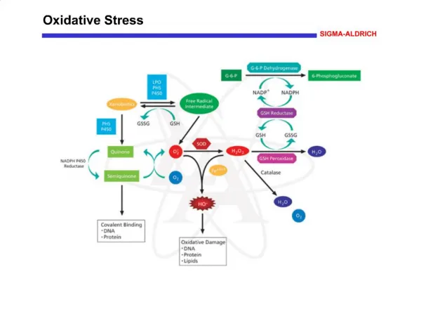
Mitochondrial Pathway Death Receptor Pathway ceramide others oxidants FasL Fas/Apo1 /CD95 DNA damage D D Bcl-2 D D D FADD DISC Procaspase 8 Apaf -1 BID Procaspase 9 Cytochrome c Caspase 8 Procaspase 3 Apaf -1 dATP dATP Caspase 3 Cellular targets apoptosome Caspase 9 Major Apoptotic Pathways in Mammalian Cells Hengartner, M.O. 2000. Nature. 407:770. Green, D. and Kroemer, G. 1998. Trends Cell Biol.8:267. Apoptosis Oxygen Society Education Program Tome & Briehl 3
Apoptotic Pathways Effectors and Modulators • There are two major apoptotic pathways in mammalian cells. • The death receptor pathway, exemplified by FasL binding to an extracellular receptor, causes the formation of the DISC that results in the activation of caspase-8. • The mitochondrial pathway is activated by most cellular stresses. A resulting signal or intracellular change causes the release of cytochrome c into the cytosol. Cytochrome c binds to Apaf-1 and procaspase-9 to form the apoptosome and catalyzes the activation of caspase-9. • Initiator caspases, such as 8 and 9, activate effector caspases that cleave multiple cellular proteins. Caspases are characterized by an active site cysteine. Further discussion on caspases can be found in Earnshaw, W.C. et al. 1999. Annu. Rev. Biochem.68:383. • Bcl-2 is a proto-oncogene that was first discovered in B-cell lymphoma. Bcl-2 prevents apoptosis by blocking the release of cytochrome c from the mitochondrion by an unknown mechanism. Several models are discussed in Hengartner, M.O. 2000. Nature407:770. There are many Bcl-2 homologs, some with pro- and others with anti-apoptotic functions. The ratio between these two types helps determine the fate of the cell. Additional information about Bcl-2 family members can be found in Gross, A. et al. 1999. Genes & Dev. 13:1899. Apoptosis Oxygen Society Education Program Tome & Briehl 4
Phagocyte ScavengerReceptors ? Oxidized LDL-like Site PS ApoptoticCell Phosphatidyl-serineReceptors C1qBindingSite C1q Bridge RAC-1 DOCK 180 C1q Receptor ELMO CRKII CytoskeletalReorganization forEngulfment Apoptosis and Phagocytosis • Phagocytes recognize “eat-me” or cell corpse signals on the apoptotic cell surface. These signal the phagocyte to activate cellular engulfment machinery. • Phosphatidylserine exposure on the target cell surface and the phosphatidylserine receptor on the phagocyte are essential for phagocytosis. • Defining other receptors, bridge molecules, “eat-me” signals and signaling molecules involved in initiating the cytosolic changes needed for engulfment are very active areas of research. The articles listed below review current knowledge and are the sources for this diagram. Savill, J. and Fadok, V. 2000. Nature. 407:784. Canradt, B. 2002. Nature Cell Biol.4:E139. Apoptosis Oxygen Society Education Program Tome & Briehl 5
1. Phagocyte ApoptoticCell With "eat me"signals 2. Phagocyte HealthyCell Phagocyte inducesapoptotic machineryin healthy cell 3. Engulfment Phagocyte Apoptotic cell inducesphagocytic machineryin phagocyte ApoptoticCell With "eat me"signals 4. PhagocytePrecursor Apoptotic cell inducesmaturation of precursorinto phagocyte ApoptoticCell With "eat me"signals Apoptosis and Phagocytosis • The first pathway shows the engulfment of an apoptotic cell exposing “eat-me” signals. • Data from mammalian systems and genetic studies from Caenorhabditis elegans have shown that phagocytes and target cells have several types of interactions. • Conradt has proposed several models (2-4) to indicate the more complex phagocyte-target interactions. Conradt, B. 2002. Nature Cell Biol. 4:E139. Greene, D.R. and Beere, H.M. 2001. Nature. 412:133. Apoptosis Oxygen Society Education Program Tome & Briehl 6
Apoptosis and Cellular Redox Environment • Oxidants such as hydrogen peroxide can trigger apoptosis. • Intracellular ROS generation by chemotherapeutics and ionizing radiation may be critical to induction of apoptosis by these agents. • Depletion of glutathione pools occurs during apoptosis and GSH depletion can increase apoptosis, in some systems. • Antioxidant enzymes and chemical antioxidants can protect against apoptosis. • Oxidative damage to lipids and DNA is seen during apoptosis in some systems. • ROS production can also attenuate apoptosis. Chandra, J. et al. 2000. Free Rad. Biol. Med.29:323. Buttke, T.M. and Sandstrom, P.A. 1995. Free Rad. Res.22:389. Apoptosis Oxygen Society Education Program Tome & Briehl 7
Apoptosis Signaling and Cellular Redox Environment • Themes emerging from research on signaling pathways include: • The activity of multiple apoptosis regulators is modulated by the cellular redox environment. Examples include p53, NF-kappaB and the JNK/SAPK and apoptosis signal-regulating (ASK1) kinases. • Downstream targets of these regulators function in the control of the cellular redox environment. Examples include differential regulation of oxidative stress-related genes during p53-induced apoptosis, regulation of the mitochondrial antioxidant protein (MnSOD) by NF-κB and phosphorylation of Bcl-2 downstream of the ASK1signaling pathway. • Whether or not the cellular redox environment is maintained in balance, following an apoptotic signal, influences the decision of cell fate: life vs. death. Sun, Y. and Oberley, L.W. 1996. Free Rad. Biol. Med.21:335. Trinei, M. et al. 2002. Oncogene21:3872. Apoptosis Oxygen Society Education Program Tome & Briehl 8
Bcl-2 and Cellular Redox Environment • Bcl-2 affects the redox environment of the cell. • Many Bcl-2 expressing cells have increased GSH. • bcl-2 knockout mice show increased oxidative stress. • Some Bcl-2-overexpressing cells exhibit increased baseline intracellular ROS (i.e., Bcl-2 acts as a pro-oxidant). • The impact of Bcl-2 on the redox environment of the cell could affect redox-sensitive transcription factors and ROS-based signaling pathways involved in apoptosis. Hengartner, M.O. 2000. Nature. 407:770. Voehringer, D.W. and Meyn, R.E. 2000. Antioxidants & Redox Signalling.2:537. Apoptosis Oxygen Society Education Program Tome & Briehl 9
Cytochrome c and Cellular Redox Environment • Cytochrome c in solution can act as an antioxidant and an ROS scavenging function for cytochrome c in the intermembrane space has been proposed by Skulachev. • Release of cytochrome c into the cytosol from the mitochondrion interrupts the electron transport chain resulting in increased production of superoxide from the mitochondrion. • Binding of cytochrome c to form the apoptosome and activate caspase-9 does not appear to depend on the ability of cytochrome c to transfer or accept electrons. • However, the reduction state of cytochrome c may still be important because reduction and oxidation cause conformational changes that may be critical for cytochrome c binding to Apaf-1 and procaspase-9. Cai, J. and Jones, D.P. 1998. J. Biol. Chem.273:11401. Hancock, J.T. et al. 2001. Free Rad. Biol. Med.31:697. Skulachev, V.P. 1998. FEBS Lett. 423:275. Apoptosis Oxygen Society Education Program Tome & Briehl 10
Caspases and Cellular Redox Environment • The cysteine in the caspase active site is sensitive to oxidation or to thiol alkylation. • Intracellular superoxide or hydrogen peroxide concentrations have been implicated in regulating caspase activity and modulating apoptosis. • The cysteine sulfhydryl can also be S-nitrosylated, inactivating the caspase and providing a mechanism to reversibly modulate caspase activity. • In some systems, caspases play a role in the life and death decision; therefore, their inactivation may promote functional cell survival. Chandra, J. et al. 2000. Free Rad. Biol. Med.29:323. Pervaiz, S. and Clement, M.-V. 2002. Biochem. Biophys. Res. Comm.290:1145. Green, D.R. and Kroemer, G. 1998. Trends Cell Biol.8:267. Apoptosis Oxygen Society Education Program Tome & Briehl 11
Oxidative Stress and Phagocytosis in Apoptosis • ROS released by macrophages can induce apoptosis in target cells suggesting a role for phagocytes in cell population control. • Phosphatidylserine is selectively oxidized in some cells in response to oxidants. • Work by Kagan and colleagues has suggested that externalization of oxidized phosphatidylserine during apoptosis may increase phagocytosis of these cells. Duffield, J.S. et al. 2000. J. Immunol.164:2110. Fabisiak, J.P. et al. 1997. Am. J. Physiol.272:C675. Apoptosis Oxygen Society Education Program Tome & Briehl 12
Insensitivity to Anti-growth Signals Self-sufficiency inGrowth Signals LimitlessReplicativePotential EvadingApoptosis Cancer SustainedAngiogenesis Tissue Invasion and Metastasis Apoptosis and Cancer Hanahan and Weinberg have proposed that normal cells must acquire six phenotypes to become malignant. One of these traits is resistance to apoptosis. In this model, the chronological order and mechanism by which these phenotypes are acquired may differ in each tumor. Genomic instability provides the driving force for acquiring new phenotypes. Thus, mutations in genomic “caretaker” systems such as p53 may increase the rate at which other alterations occur. Hanahan, D. and Weinberg, R.A. 2000. Cell. 100:57. Apoptosis Oxygen Society Education Program Tome & Briehl 13
Oxidative Stress and Cancer Disorders Sharing Oxidative Stress and Cancer Proneness In each of these congenital disorders the cells show evidence of increased oxidative stress. Affected individuals show an increased incidence of cancer. Chromosomal instability is also a common feature of the first four disorders. Taken together these data suggest that the increased oxidative stress may contribute to development of genomic instability (a mutator phenotype) that is a hallmark of cancer cells. Franconi anaemia Xeroderma pigmentosum Ataxia telangiectasia Bloom syndrome Down syndrome Cystic fibrosis Pagano, G. et al. 1998. Medical Hypotheses. 51. 253. Loeb, L. 2001. Cancer Res.61:3230. Apoptosis Oxygen Society Education Program Tome & Briehl 14
Oxidative Stress and Cancer Chronic Inflammation is Associated with Malignancy Apoptosis Oxygen Society Education Program Tome & Briehl 15
· Neutrophil · · Eosinophil · Macrophage Oxidative Stress and Cancer Oxidants Released byInflammatory Cells O2- H2O2 HOCl NO ONOO- NO2 HO 1O2 The continuous production of oxidants at the site of chronic inflammation may cause cancer. Fitzpatrick, F.A., 2001. Int. Immunopharmacol. 1:1651. O'Byrne, K.J., and Dalgleish, A.G., 2001. Br. J. Cancer, 85:473. Kuper, H., et al. 2000. J. Int. Med., 248:171. Shacter, E., and Weitzman, S.A., 2002. Oncology16:217. Apoptosis Oxygen Society Education Program Tome & Briehl 16
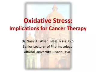
Anti-Cancer Agents Doxorubicin Daunorubicin Mitomycin C Etoposide Cisplatin Arsenic trioxide Ionizing radiation Photodynamic therapy Oxidative Stress and Cancer Therapy These anti-cancer agents depend exclusively or in part on the production of reactive oxygen species for cytotoxicity. Sensitivity of tumor cells to oxidative stress and/or apoptosis may affect treatment success. Davis, W. 2001. Pharmacol. Exp. Ther.296:1. Apoptosis Oxygen Society Education Program Tome & Briehl 17
40 30 % Apoptosis 20 10 0 Neo3 THX CAT38 Cell Variant Oxidative Stress, Apoptosis and Cancer WEHI7.2 mouse thymoma cells overexpressing catalase (CAT38) or thioredoxin (THX) are resistant to glucocorticoid-induced apoptosis in vitro. This suggests that glucocorticoids induce apoptosis by an ROS-dependent mechanism. Percentage of annexin positive, propidium iodide negative cells in the culture after a 24h treatment with glucocorticoid. Tome, M.E. et al. 2001. Cancer Res. 61:2766. Tome, M.E. and Briehl, M.M. 2001. Cell Death Differ. 8:953. Apoptosis Oxygen Society Education Program Tome & Briehl 18
Oxidative Stress, Apoptosis and Cancer Average tumor weight is increased in SCID mouse tumor xenografts from cells overexpressing catalase or thioredoxin. Tumors from both transfectants contain fewer apoptotic cells, but mitotic cell numbers are similar. This suggests that antioxidant overexpression results in increased tumor size due to a decrease in apoptosis. Values are means ± SEM. *Indicates the number of apoptotic cells in a 100 X microscope field. Apoptosis Oxygen Society Education Program Tome & Briehl 19
Carcinogenesis Antioxidants ? Chronic Oxidative Stress Genetic instability Tumor Progression New phenotypes Apoptosis Growth Etc. inflammation genetic disorder environment Cancer Therapy Oxidative Stress, Apoptosis and Cancer: Some Models Tumor Regression NO ROS-based Therapy Tumor contains apoptosis- or oxidant-resistant cells Tumor Tumor Progression YES Antioxidants ? Apoptosis Oxygen Society Education Program Tome & Briehl 20