
ABSTRACT
Treating Allergic Conjunctivitis Induced in BALB/c Mice. Ami Casis, Dr. Steven Spilatro, Marietta College. RESULTS. ABSTRACT

ABSTRACT
E N D
Presentation Transcript
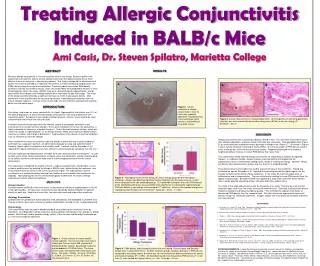
Treating Allergic Conjunctivitis Induced in BALB/c Mice Ami Casis, Dr. Steven Spilatro, Marietta College RESULTS ABSTRACT Seasonal allergic conjunctivitis is the most common form of eye allergy. Based on patient self-assessments of treatment effects, clinical studies have shown that topical treatments are more effective than oral treatments in alleviating symptoms. This study investigated the effectiveness of topical Visine and oral Benedryl in reducing inflammation as measured by polymorphonuclear cell (PMN) density along the conjunctival epithelium. Previous studies have shown PMN density correlates inversely to treatment efficacy, and in this study PMNs were predicted to be lower in mice treated topically rather than orally. BALB/c mice were sensitized toward ragweed pollen, allergic conjunctivitis was induced, and histology sections were examined via light microscopy. The onset of the allergy was determined by a significant increase (p<0.05) in granulocyte density. Mice treated with Visine and Benedryl did not show significance in decreasing cell migration from the area of allergen exposure. Contrary to the clinical study, the oral treatment alleviated cell migration better than the topical agent. A A B * * INTRODUCTION Eye allergy, also known as ocular conjunctivitis, is a type I hypersensitivity that affects over 20% of the general population, of which seasonal allergic conjunctivitis is the most predominant form around the world.5 Symptoms of eye allergies include itchiness, irritation, mucus discharge, tears, and swelling of the lining of the eye and eyelid. Seasonal conjunctivitis develops when the immune system of susceptible individuals reacts excessively to an usually harmless allergen. The mucous membrane of the eye, the conjunctiva, is highly vulnerable to airborne or microbial allergens.2 One of the most common allergen, which was used in this study, is ragweed pollen. In an allergic reaction, PMNs show significant degranulation, increase in number, and change in distribution.4 Degranulation is the release of chemical mediators such as histamine to rid of the allergen. Most treatments for ocular conjunctivitis target the allergic reaction after the onset of symptoms.3 Antihistamines, a popular treatment, are administered topically or orally and sometimes both.3 Recently, topical agents have become more widely used.3 A clinical study by Alexander et. al.1 reported that topical antihistamine was more efficient in alleviating allergic symptoms than the oral.1 A murine model was developed to best resemble the human disease and to test therapies.7 In order to make sense of the clinical assessments of topical versus oral treatments, a better understanding of the cellular activities in the eye will help relate the immunologic processes with the clinical observations. This experiment investigated the effects of Visine, a topical vasoconstrictor, and Benedryl, an oral systemic antihistamine, by inducing the allergy in BALB/c mice, administering the treatments, and evaluating the levels of immune cells in the conjunctivial region. The subcutaneous route of sensitization is a modified technique derived from traditional murine models that injected into the footpads. Total lymphocytes were examined histologically to determine the onset of allergic conjunctivitis and the effects of treatments. * Figure 2. Clinical symptoms of allergic conjunctivitis in mice 24 hours after ragweed application included tearing, mucus discharge (white asterisk), and swelling of the eyelids (arrow). Figure 5. Cellular observations of enlarged goblet cells. (A) Enlarged mucin-secreting goblet cells (asterisk) was more prevalently observed in allergy mice (B) than in the non-allergy (C). Scale bar = 50 m. DISCUSSION Allergic conjunctivitis was successfully induced in BALB/c mice. The mice were sacrificed 24 hours after allergen application, and peak cellular infiltration was evaluated.7 Enlarged goblet cells (Figure 5) are associated with heightened mucus discharge in allergic mice (Figure 2).7 As shown in Figures 3 and 4, cellular infiltration consisted of mostly PMNs. The inverse relation of PMN density and the onset of allergy was similar to that of previous studies. Ragweed challenged mice showed a heightened cellular increase in overall PMN density than naive mice.7 This experimental model showed allergic conjunctivitis via subcutaneous injection of ragweed allergen. In traditional models, allergen injection was inserted into the footpad, but the subcutaneous route of sensitization yielded similar results in inducing the allergy. However, allergy cell density from subcutaneous injection was less than in the footpad method.7 The subcutaneous murine model was used to evaluate efficacy of two treatments. Testing two antihistamine agents, Alexander et al.1 reported that patients preferred the topical agent over the oral due to faster-acting relief of allergy symptoms. In this study, the treatment types were an antihistamine and a vasoconstrictor. The oral antihistamine resulted in lesser PMN density than the topical vasoconstrictor. Benadryl-treated mice appeared to show fewer cells than Visine-treated mice, thereby possibly alleviating cellular infiltration better than topical Visine. The result of this study did not concur with the Alexander et al. study. This may be a result of the treatment types rather than the routes of treatment administration. Generally, antihistamines bind to histamine receptors and reduce symptoms quickly.1 The antihistamine appears to be better at decreasing PMN density than the vasoconstrictor. Still, both treatments appeared to reduce cell density, though not statistically significant. A bigger sample size may decrease variability and effect size measurement. C A B Figure 3. Histological results of non-allergy (A) versus allergy group (B) after the topical challenge. Allergy mice did show significant cellular migration into the conjunctiva (p < 0.031) when compared to the non-challenged group. (C) Independent sample t-test of the PMN density of the standard counting area was used determine significance in inducing the ragweed allergy. PMN cell density in non-allergy mice averaged 2.77 cells/mm2, whereas the ragweed allergy mice showed elevated numbers of 6.07 cells/mm2. Scale bar = 50m. r2 = 83%. METHODS Allergy Induction Female 8-weeks-old BALB/c mice were given a subcutaneous injection of ragweed pollen in ALUM in the right shoulder. Ten days later, conjunctivitis was induced by topical instillation of ragweed directly on each eye. Twenty-four hours later, the mice were sacrificed under ether anesthesia. Histology Examination Eyeballs were enucleated with eyelid attached, fixed, dehydrated, and embedded in LR White resin. Tissues sections (3μm) were mounted on gelatin-coated slides, stained (H & E), and photographed. Data Analysis Cells (total PMNs) were counted in blinded standard areas adjoining the conjuctival fornix by volunteers. An independent sample t-test was used to compare cell density of non-allergy vs. allergy groups. ANOVA was used to compare allergy control, Visine-treated, and Benedryl-treated groups. p < 0.05 was considered significant. • REFERENCES • 1. Alexander M., L. Rosen, W. Yang. 1999. Comparison of topical nedocromil sodium and oral terfenadine for the treatment of seasonal allergic conjunctivitis. Clinical Therapeutics. 21(11): 1900-1907. • 2. Bisca M. 1997. Current therapy of allergic conjunctivitis. Current Therapeutic Research. 58(11): 828-841. • Bielory, L. 2002. Ocular allergy guideline: a practical treatment algorithm. Therapy in Practice. 62(11): 1611-1634. • 4. Bonini S. A., Lambiase, S. Bonini. Genetic and Environmental Factors in Ocular Allergy. Mark Abelson. Allergic Diseases of the Eye. W.B. Saunders Company. Philadelphia. 260pp. • 5. Dogru M., A. Ozmen, H. Erturk, O. Sanli, A. Karatas. 2002. Changes in tear function and the ocular surface after topical olopatadine treatment for allergic conjunctivitis: an open-label study. Clinical therapeutics. 24(8): 1309-1321. • 6. Groneberg, D.A., L. Bielory, A. Fischer, S. Bonini, U. Wahn. 2003. Animal models of allergic and inflammatory conjunctivitis. Allergy. 58: 1101-1113. • 7. Magone M.T., C. Chan, L.V. Rizzo, A.T. Kozhich, S.M. Whitcup. 1998. A novel murine model of allergic conjunctivitis. Clinical Immunology and Immunopathology. 87(1): 75-84. • 8. Magone M.T., Whitcup S.M., Fukushima A., Chan C., Silver P.B., Rizzo L.V. 2000. The role of IL-12 in the induction of late-phase cellular infiltration in a murine model of allergic conjunctivitis. Journal of allergy Clinical Immunology. 105(2): 299-307. B A C Figure 1. Gross anatomy of mouse eyeball viewed sagittally. The tissue sample was fixed in Histochoice Tissue Fixative MB, washed with phosphate buffer solution, dehydrated in ethanol, and embedded in LR White Resin. The tissue section (3μm) was stained with Hemotoxylin and Eosin. The scale = 200μm. (A) Lens (B) Retina (C) Eyelid (D) Cornea (E) Iris (F) Sclera (G) Conjunctival Fornix Figure 4. PMN density and histological sections of non-treated, Visine-treated, and Benadryl-treated mice in conjunctival fornix. (A) Both treatment groups showed a decrease in PMN density compared to the allergy control, but there was no overall significant difference between the control and treatment groups (P = 0.093). (B) Benedryl-treated mice showed less PMN density (P = 0.08) than (C) mice treated with topical Visine ( p = .63). Scale bar = 50 m. ACKNOWLEDGEMENTS: I would like to thank my principal research advisor, Dr. Spilatro, and capstone instructor, Dr. Hogan of the Marietta College Biology Department. I would like thank Dr. McShaffrey for his assistance in imaging. I would like to thank Dr. Reisner, O.D., for the capstone idea and Dr. Jones, M.D., for the surgical advice. Thank you Andrea Marion, Stacey Burgess, Jessica Craig, Toni Stephenson, and Craig Meredith for counting cells. I would like to acknowledge the Marietta College Investigative Studies Program for their support of my undergraduate research.


