Download
1 / 38
390 likes | 780 Vues
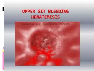
Dr. Salem Mohammad Bazarah MD, M.Ed, FACP, FRCPC, FRCPC (GI) & PhD . Management of a Pt with Hematemesis. A common medical condition. 250,000 – 500,000 admissions/year US UGI bleeding incidence 100/100,000 adults Incidence increases 20-30 fold from third to ninth decade of life

E N D
Dr. Salem Mohammad Bazarah MD, M.Ed, FACP, FRCPC, FRCPC (GI) & PhD Management of a Pt with Hematemesis
A common medical condition • 250,000 – 500,000 admissions/year US • UGI bleeding incidence 100/100,000 adults • Incidence increases 20-30 fold from third to ninth decade of life • LGI bleeding incidence 20/100,000 adults • Overwhelmingly disease of the elderly • GI bleeding stops spontaneously in 80 %
Morbidity Data • Majority will receive blood transfusions • 2 – 10 % require urgent surgery to arrest bleeding • Average LOS 4 – 7 days • Mortality rates for UGI bleeding 2 – 15 % • Mortality for patients who develop bleeding after admission to hospital for another reason is 20 – 30 %
Costs • Average hospital costs exceed $ 5,000 per admission • Most of this for hospital bed and ICU stays rather than physician fees, blood products, diagnostic tests, or medications • Reduction of hospital admissions and LOS has greatest potential to reduce costs
UGI bleeding:Nomenclature • Hematemesis 25 % • Melena alone 25 %, 50 – 100 cc of blood will render stool melenic • Hematochezia 15 %, seen in massive UGI hemorrhage • “Red blood” hematemesis • “Coffee ground” emesis
Indications for Hospitalization and Intensive Care • Traditional: Endoscopy on the day of admission or on the day after • Recent studies: Complete endoscopic risk stratification PRIOR to admission • Between 25- 30 % of patients with UGI bleeding could be discharged from the Emergency Department
History • 45 yrs male with 1 day hx of vomiting blood
Approach • Assess the severity • Resuscitate • Establish the site of bleeding • Endoscopic intervention • Reassess severity: liase with surgical team • Medical treatment • Indications for surgery
Assessing severity: Rockall criteria CriterionScore • Age <60 years 0 60-79 yrs 1 >80 years 2 • Shock None 0 Pulse & sBP >100 1 sBP <100 2 • Co-morbidity None 0 Cardiac/any major 2 Renal/liver/malig. 3 • Total initial score (max = 7)
Implications of initial score Initial risk score (pre-endoscopy) ScoreMortality 0 0.2% 1 2.4% 2 5.6% 3 11.0% 4 24.6% 5 39.6% 6 48.9% 7 50.0% Rockall TA et al Gut 1996; 38: 316-21
Resuscitate • Large bore intravenous cannula x 2 • X-match 4 units, give colloid & transfuse if • Fresh melaena on PR • Postural hypotension >15mm/Hg • sBP <100mmHg • Cross match 6 units for • Suspected variceal bleeding • Otherwise group and save serum only
Resuscitation • Indications for CVP • Rockall score > 3, first rebleed, or inadequate access • Insert urinary catheter if CVP appropriate • Urea/creatinine ratio • If >unity (eg 12.4/90), then upper GI bleed likely • Monitor Pulse & BP ‘?hrly’ • Guide of halves: if pulse higher or BP lower than last recording, then halve the time to the next recording • If pulse trend rises on 3 occasions, call senior cover
Establish site of bleeding • Endoscopy on next available list • Ideally <24hr • Out of hours endoscopy • If a surgical decision depends on the result • Therefore consent ‘endoscopy, ?proceed’ • Check endoscopy report for • stigmata of recent haemorrhage • intervention
Stigmata of recent haemorrhage • Clean ulcer base (rebleed <1%) • Black spots ulcer base (rebleed 5%)
Stigmata of recent haemorrhage • Fresh clot (rebleed 30%) • Visible vessel (rebleed 50%)
Stigmata of recent haemorrhage • Bleeding vessel (rebleed 80%)
Upper GI Bleeding Klaus Gottlieb, MD, FACP, FACG
Source of bleeding Common • DU (35%) • GU (20%) • Oesophagitis (6%) • Mallory-Weiss (6%) • No source found (20%) Uncommon/Rare • Varices • Tumour • Aortoenteric fistula • Dieulafoy • Haemobilia • Angiodysplasia
Intervention • Endoscopic injection with • Adrenaline 1:10 000, thrombin, sclerosant, or saline all halve the risk of rebleeding • As good as heater probe, laser therapy • Tranexamic acid • 1g iv three times daily for 72hr reduces mortality • Omeprazole 60mg iv stat and infusion 8mg/hr for 72hr • may reduce mortality after endoscopic intervention • Nothing else has been shown to work Do not prescribe iv ranitidine, or oral PPI until after endoscopy
Reassess severity: update Rockall Score • Endoscopic diagnosis • No lesion, or M-W tear 0 • All other diagnoses 1 • Malignancy of upper GI tract 2 • Stigmata of recent haemorrhage • None/haematin 0 • Clot, visible vessel,blood in stomach 2 • Final score after endoscopy(max 11)
Updated Rockall score Initial score (pre-endoscopy) ScoreMortality 0 0.2% 1 2.4% 2 5.6% 3 11.0% 4 24.6% 5 39.6% 6 48.9% 7 50.0% Final score (after endoscopy) ScoreMortality 0 0% 1 0% 2 0.2% 3 2.9% 4 5.3% 5 10.8% 6 27.0% 7 17.3 8+ 41.1%
Further management • Liase with surgeons if • Initial score >3 (ie if CVP necessary) • Posterior duodenal ulcer • Final Rockall score >4 • After endoscopy • Eat & drink if no stigmata, or haematin only • Clear fluids for 12 hr if endoscopic intervention • NBM only if haemostasis not secure (varices) • Re-examine after 4-8hr for signs rebleeding • Ring blood bank to keep blood available for 24hr after endoscopic intervention
Signs of rebleeding • Rise in pulse rate • Fall in CVP • Decrease in hourly urine output • Further haematemesis or fresh melaena • Look at the patient as well as the charts! • Act if rebleeding suspected • FBC and transfuse • Ensure large bore access, central line and catheter • Call surgical team
Indications for surgery • Early surgery (esp. elderly) assoc. with lower mortality • Age over 60 years • Transfusion >4 units in 24hr • One rebleed • Continued bleeding • Age under 60 years • Transfusion >8 units in 24hr • Two rebleeds • Continued bleeding • Decision not to operate should be taken by consultant
Special notes - Variceal bleeding • Suspect variceal bleeding if…..- Alcohol Hx- Deranged LFT’s- Jaundice*- Hyponatraemia*- Ascites*- Coagulopathy- Low platelets- Previous Hx of varices*
Special notes – Variceal Bleeding • Resuscitate • Correct coagulopathy (FFP x 4 and vit K IV) • Endoscopy andbanding/sclerotherapy • Glypressin 2mg iv stat and 1-2mg repeated 4hrly • Treatother aspects of decompensation • Ascites (spironolactone, no N/saline) • Encephalopathy (lactulose, no sedation) • Renal impairment (avoid hypovolaemia) • Malnutrition (iv vitamins, fine bore feeding) • Underlying liver disease (hepatic ‘screen’, aFP etc) • Post-bleed prophylaxis
Summary • Objective assessment (Rockall criteria) • Resuscitation before endoscopy • Monitor by rule of halves: look for trends • No role for empirical acid suppression • Critical appraisal of endoscopy report • Liaise with surgeons early • Discriminate between high & low risk patients