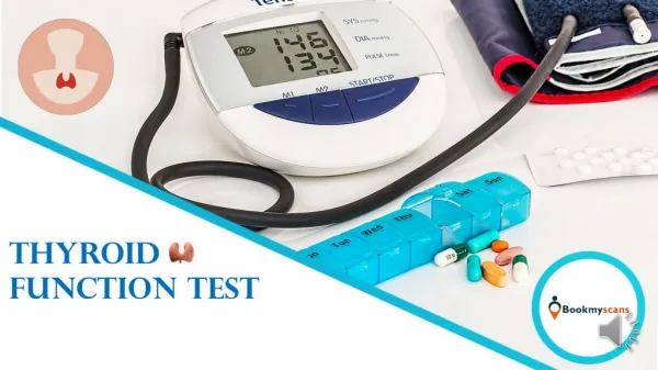
Thyroid Function Tests
Thyroid Function Tests. Hasan AYDIN, MD Yeditepe University Medical Faculty Endocrinology and Metabolism. Tests of Thyroid Function:. Tests of thyroid hormones in the blood Evaluation of hypothalamo-pituitary-thyroid axis Assessment of iodine metabolism Estimation of gland size

Thyroid Function Tests
E N D
Presentation Transcript
Thyroid Function Tests Hasan AYDIN, MD Yeditepe University Medical Faculty Endocrinology and Metabolism
Tests of Thyroid Function: • Tests of thyroid hormones in the blood • Evaluation of hypothalamo-pituitary-thyroid axis • Assessment of iodine metabolism • Estimation of gland size • Thyroid biopsy • Observation of the effects thyroid hormones on peripheral tissues • Measurement of thyroid autoantibodies
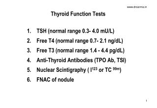
Tests of Thyroid Hormones in Blood • Total serum T4 • Free T 4 level • Total serum T3 • FT4-I • FT3 and rT3 • Thyroglobulin
Total T3 and T4 • Protein bound + free hormone • Measured by radioimmunoassay(RIA) or immunufluorescent assay. • Provide reliable index of thyroid gland activity if levels of thyroid binding proteins normal. • Changes in serum concentration of thyroid-binding proteins(TBP) or the presence of drugs that effects binding of T3 and T4 to TBP will modify total hormone levels, but not the biologically active free T3 and T4 levels.
Total T3 (Normal: 95-190 ng/dl) • The T3 is increased in almost all cases of hyperthyroidism and usually goes up before the T4 does. • T3 is a more sensitive indicator of hyperthyroidism than the Total T4. • In hypothyroidism the T3 is often normal even when the T4 is low. • The T3 is decreased during acute illness and starvation, and is affected by several medications including beta blocker, steroids and amiodarone.
Total T4 (Normal: 5-12 ng/dl) • In circulation most T4 (99.8%) is bound to specific plasma proteins. TBG (60-75%),Prealbumin/transthyretin 15-30%, albumin 10% • In the absence of thyroid dysfunction, abnormal results are usually due to an abnormal level of serum TBG. • The TT4 level may not always correspond to the FT4 concentration • Free hormone is only 0.03 % of the total.
Factors Influencing Concentration of Protein Bound Thyroid Hormones A- Increased TBG concentration Congenital Hyperestrogenic states: Pregnancy, Estrogen therapy Diseases: Acute infectious hepatitis, hypothyroidism B- Decreased TBG concentration Congenital Drugs: Androgenic steroids, glucocorticoids Major systemic illness: Protein malnutrition, nephrotic syndrome, cirrhosis, hyperthyroidism C- Drugs affecting thyroid hormone binding to TBG Phenytoin Salycilates Diazepam......
Free T3 and Free T4 Free T3 (N: 0.2 - 0.52 ng/dl) and T4 hormone levels can be measured directly by dialysis method by chemiluminescent assay estimated using free thyroxine index • Free T4 (Normal: 0.9 - 2 ng/dl) • The FT4 measures the concentration of free thyroxine, the only biologically active fraction in the serum. • The free thyroxine is not affected by changes in concentrations of binding proteins Thus such conditions as pregnancy, or estrogen and androgen therapy do not affect the FT4.
Thyroid Stimulating Hormone • A glycoprotein secreted bythe anterior pituitary • Structurally similar to FSH, LH and chorionic gonadotropin (hCG) • Composed of two dissimilar non-covalently bound α-subunit andβ-subunits • The α-subunit is hormone non-specific • The β-subunit is distinct for each hormone and confers biological andimmunological specificity • Antibodies used in immunoassays of TSH target the β-subunit
Thyroid Stimulating Hormone • Measures the response of the pituitary gland to circulating T4 and T3. • Elevated inhypothyroidism due to primary thyroid disease, iodine deficient goiter. • In hyperthyroidism, TSH will be suppressed. • This is probably the most important test that can be performed to assess thyroid function. • Highly sensitive TSH test is available. This method utilizes monoclonal antibodies directed against the beta subunit of the molecule and is therefore specific for TSH.
The effect of serum TSH assay sensitivity on the discrimination of euthyroid subject (Euth) from those with thyrotoxicosis
Measurement of TSH Methods Principle Comments Radioimmunoassay Competitive binding of (RIA) radiolabeled TSH and non- Currently being labeled TSH to limited binding phased out sites on antibody Immunoradiometric Binding of TSH to radiolabeled Most sensitive assay antibody and widely used ELISA Binding of TSH to enzyme- labeled antibody ILMA Binding of TSH to luminescent labeled-labeled antibody Sensitive
Evaluation of Hypothalamo-Pituitary-Thyroid Axis • The integrity of the hypothalamo-pituitary-thyroid axis can be evaluated by • the response of the pituitary gland to thyroid hormone excess or deficiency; • the ability of the thyroid gland to respond to thyrotropin (TSH); • the pituitary responsiveness to thyrotropin-releasing hormone (TRH). • These tests are intended to identify the primary organ affected by the disease process that manifests as thyroid dysfunction; primary (thyroid), secondary (pituitary), or tertiary (hypothalamic) malfunction.
TRH Test • Procedure:IV injection of • TRH 100 - 200 µg.Time-course measurement of TSH-15, 0, 15 and 30 mn. examines interrelationship of TRH and TSH secretionsby administering exogenous TRH
Clinical Utility of Thyroid-Related Laboratory Tests Name of Test Abbreviation Clinical Utility Tests for Evaluation of Thyroid Status Thyrotropin (sensitive IA) sTSH Best general test (Conventional RIA) TSH phased out Free thyroxine (appropriate method) FT4 Second best general test Free (3,5,3') triiodothyronine FT3 Adjunct test, diagnosis of T3 toxicosis Total (3,5,3') triiodothyronine T3 Used in lieu of FT3 Total thyroxine T4 Inadequate as general test Thyroglobulin Tg Valuable in thyroid cancer Free T4 index FT4/FTI T4 x T3-BR Replaced by FT4 T4 x T4-BR
Thyroglobulin (Tg) • In serum by double antibody immunoassay • The normal range vary with method and laboratory , • Normal range is less than 40 ng/mL ( < 40 mikrogram/L) in the euthyroid individual • <5 ng/mL in a thyroidectomized individual. • The major problem endogenous Tg Ab • Spuriously low or spuriously high values.
Thyroglobulin (Tg) Elevations • Graves ' disease • Toxic MNG • Acute or chronic thyroiditis • Papillary or follicular thyroid carcinoma. • Following thyroidectomy and 131-I therapy, • serum thyroglobulin greater than 10 ng/dL (> 10 ~g/L) indicates the presence of metastatic disease • rise in a patient with known metastases indicates progression of the disease.
Thyroid Autoantibodies Thyroid peroxidase (TPO) (or antimicrosomal (TMAb) and antithyroglobulin (TgAb) antibodiesare measured to confirm or rule out autoimmune thyroid disease. High titers of these antibodies are present in 97 % of patients with Grave’s disease or Hashimoto’s thyroiditis. Not specific for the type of disease. Generally these are measured using RIA, ELISA or IRMA techniques Reported prevalence of thyroid antibodies in population is highly variable ( %2-30) Thyroid receptor stimulating antibody is characteristic of Grave’s disease (Present in 90%). Helpful in diagnosis of euthyroid Grave’s ophtalmopathy and in predicting possible occurrence of thyroid dysfunction in neonates Thyroid receptor blocking antibody is characteristic of autoimmune thyroid disease
Thyroid peroxidase (TPO) and Antithyroglobulin (TgAb) • Measured to confirm or rule out autoimmune thyroid disease. • High titers of these antibodies are present in 97 % of patients with Grave’s disease or Hashimoto’s thyroiditis. • Not specific for the type of disease. • Generally these are measured using RIA, ELISA or IRMA techniques • Reported prevalence of thyroid antibodies in population is highly variable (2-30%)
Iodine Metabolism & Biosynthetic Activity • Radioactive iodine uptake • RAIU supression test
RAIU • Radioactive iodine allows assessment of the turnover of iodine by the thyroid gland in vivo. • Iodine-123 is the ideal isotope for this purpose • Administered orally at a dose of 100-200 micCi, • Radioactivity over the thyroid area is measured with a scintillation counter at 4 or 6 hours and again at 24 hours
RAIU • Vary with the iodide intake. • In areas of low iodide intake and endemic goiter, the 24-hour RAIU may be as high as 60-90%. • The normal uptake at 6 hours is 5-15% and at 24 hours 8-30%. • In thyrotoxicosis due to Graves' disease or toxic nodular goiter, the 24- hour RAIU is markedly elevated
RAIU Supression Test • 75-100 microgram of T3 in divided doses daily for 5 days • Reduce the 24-hour RAIU by more than 50% (suppression test). • Failure of the thyroid to suppress on this treatment indicates autonomous thyroid function, as in Graves' disease, or autonomously functioning thyroid nodules.
Thyrotoxicosis with a Very Low Thyroidal RAIU • Subacute thyroiditis • During the active phase of Hashimoto's thyroiditis, • Thyrotoxicosis factitia due to oral ingestion of a large amount of thyroid hormone • As a result of excess iodide intake (amiodarone therapy), inducing thyrotoxicosis in a patient with latent Graves ' disease or multinodular goiter, the low uptake being due to the huge iodide pool; • Struma ovarii • Ectopic functioning metastatic thyroid carcinoma after totalthyroidectomy.
Thyroid Imaging • Radionuclide imaging • Radioisotope Scanning • Fluorescent Scanning • Thyroid Ultrasonography • Magnetic Resonance Imaging
Radionuclide Imaging • 123-Iodine oraIly in dose of 200-300 microCi, scanof the thyroid at 8-24 hours • TechnetiumTc 99 m pertechnetate (99mTc as 4) intravenously in a dose of 1-10 mCiscan is obtained at 30-60 minutes. • Images can be obtained either a rectilinear scanner or a gamma camera.
Radionuclide Imaging • Both the size and shape of the thyroid gland • The geographic distribution of functional activity in the gland. Functioning thyroid nodules ''hot'' nodules, Non-functioning ones ''cold'' nodules. • The incidence of malignancy in hot nodules is about 1%, but they may become toxic, producing enough hormone to suppress the rest of the gland and induce thyrotoxicosis.
Fluorescent Scanning • Advantage of this procedure patient receives no radioisotope gland can be imaged even when it is loaded with iodine ( after IV contrast media) • Disadvantage requires specialized equipment that may not be generally available.
Thyroid Ultrasonography • Thyroid size and nodularity much better detail • Useful for measuring the size of the gland or individual nodules • Evaluating the results of therapy. • Differentiating solid from cystic lesions • Guide the operator to a deep nodule during fine- needle thyroid aspiration biopsy. • Limited to thyroid tissue in the neck, ie, it cannot be used for substernal lesions.
Magnetic Resonance Imaging • Thyroid size and nodularity much better detail • Excellent image of the thyroid gland, including posterior or substernal extension of a goiter or malignancy . • Both transverse and coronal images • Lymph nodes as small as 1 cm • Invaluable for the demonstration of tracheal compression from a large goiter, tracheal invasion or local extension of a thyroid malignancy , or metastases to local or mediastinal lymph nodes.
Fine-Needle Aspiration Biopsy • The best method for differentiation of benign from malignant thyroid disease. • Outpatient procedure • Requires no preparation. • The skin over the nodule is cleansed with alcohol, • If desired, a small amount of 1 % lidocaine can be injected intracutaneously for local anesthesia.
Effects of Thyroid Hormones on Peripheral Tissues • A measurement of basal oxygen in the intact organism the basal metabolic rate (BMR). • However, this test is nonspecific and insensitive and is rarely used today • Photomotogram. The traction and relaxation time of the Achilles tendon
Algorithm for Diagnosis of Thyroid Dysfunction TSH Decreased Normal Increased FreeT4&T3 Euthyroid No further testing FreeT4 DecreasedNonthyroidal illness or drug effect Secondary Hypothyr. Normal Subclin hypothyroidism IncreasedHyperthyroidism Increased Thyroid Resistance or Pituitary Tm. Normal Subclin hyperthyroidism Glucocort. Decreased Primary Hypothyroidism