Download
1 / 31
430 likes | 1.09k Vues
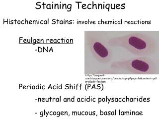
Staining Techniques Histochemical Stains: involve chemical reactions Feulgen reaction -DNA Periodic Acid Shiff (PAS) -neutral and acidic polysaccharides - glycogen, mucous, basal laminae. http://bioquant-com.bioquantusers.org/products.php?page=ls&content=gallery&sub=feulgen.

E N D
Staining Techniques • Histochemical Stains: involve chemical reactions • Feulgen reaction • -DNA • Periodic Acid Shiff (PAS) • -neutral and acidic polysaccharides • - glycogen, mucous, basal laminae http://bioquant-com.bioquantusers.org/products.php?page=ls&content=gallery&sub=feulgen
Goblet cells PAS stain Intestinal Villus
Staining Techniques • Localization (staining) of an enzyme • AB + T AT + B ENZYME provide substrate • generate visible product
Staining Techniques • AB + T AT + B • Acetylcholinesterase- neuromuscular junction ACETYL CHOLINESTERASE Other stains for ATPases, alkaline phosphatases, and others
A technique to localize specific molecules in an organ, tissue or cell. IMMUNOCYTOCHEMISTRY
An organism creates antibodies to foreign molecules, ANTIGENS. An antigen may have different regions, EPITOPES, that are recognized as foreign by an organism. First, a bit of immunology……….
Polyclonal antibodies -A collection of distinct types of antibody molecules that recognize the same antigen (antibodies A + B + C)
Monoclonal antibodies -A single type of antibody molecule that recognizes only one epitope on an antigen (antibody A OR B OR C)
Polyclonal antibodies • ADVANTAGES: recognize more epitopes in tissue • DISADVANTAGES: less specificity • Monoclonal antibodies • ADVANTAGES: more specific • DISADVANTAGES: reduced signal possible
EXPERIMENT: • Homogenize a sample of human muscle containing a variety of cells (muscle cells, neurons, capillaries, connective tissue cells). • Inject homogenate into a mouse. • WHAT HAPPENS IN THE MOUSE? • Take of sample of mouse blood, extract the serum, stain a section of human muscle. • WHAT WILL BE STAINED IN THE HUMAN MUSCLE? • HOW DO WE GET STAINING OF ONLY MUSCLE MYOSIN?
Antibody against laminin Antibodies against different epitopes of myosin heavy chain Representative myosin heavy chain (MHC) immunocytochemistry images of an emphysematous diaphragm after co-incubation with anti-laminin antibody and an antibody against one of the adult MHC isoforms.
IMMUNOCYTOCHEMISTRY Use of antibodies to detect specific molecules (antigens) in a tissue Antibody binds to an antigen in the tissue. ANTIBODY ANTIGEN
IMMUNOCYTOCHEMISTRY Direct Immunocytochemistry: a visible marker is directly attached to antibody binding the antigen The antibody is conjugated to visible marker. • Fluorochrome • Enzyme (HRP) • Electron dense molecule (ferritin, gold) Procedure: Fix the tissue Rinse with saline solution Incubate with conjugated antibody Rinse Mount on slide, view with microscope
DIRECT IMMUNOCYTOCHEMISTRY ADVANTAGES Specificity Less background staining DISADVANTAGES Low sensitivity if the antigen is present in the tissue in low concentrations. Need to directly conjugate marker to antibody.
INDIRECT IMMUNOCYTOCHEMISTRY • Primary antibody binds to the antigen.
INDIRECT IMMUNOCYTOCHEMISTRY • Primary antibody binds to the antigen. • Secondary antibody binds to the primary antibody. • Secondary antibody is conjugated to a visible marker. Procedure: Fix the tissue Rinse Incubate unlabeled primary antibody Rinse Incubate labeled secondary antibody Rinse Mount on slide, view with microscope
INDIRECT IMMUNOCYTOCHEMISTRY ADVANTAGES Amplification of the signal Can use labeled secondary with different primary antibodies DISADVANTAGES The nonspecific background may increase Takes longer to do Needs more reagents
LIMITATIONS OF IMMUNOCYTOCHEMISTRY Cross-reactivity Sensitivity Antigenicity -Frozen sections
Antibodies (immunoglobulins) of specific species are used as antigens to generate secondary antibodies. ANTIGEN--> mouse antibody Rabbit anti-mouse IgG Goat anti-mouse IgG Donkey anti-rabbit IgM
QUESTION: Dr. Reist is studying the distribution of two proteins, FasII and spectrin in neurons. She would like to label both molecules in the same sample using double-labeling immunocytochemistry. She has these antibodies: Primary antibodies: Secondary antibodies: rabbit anti-FasII mouse anti-rabbit-FITC(fluorescein) mouse anti-FasII donkey anti-rabbit-FITC goat anti-FasII rat anti-mouse-Rh (rhodamine) rat anti-spectrin goat anti-mouse-Rh rabbit anti-spectrin rabbit anti-Goat-Rh donkey anti-spectrin • What primary and secondary antibodies will successfully distinguish the distribution of FasII and spectrin in the same preparation?
AUTORADIOGRAPHY • Tissue with radiolabeled molecule • Cover with photo emulsion • Radiation activates silver -> silver grains • Develop and view http://course1.winona.edu
In situ hybridization Labeled DNA or RNA probe Why?
In situ hybridization Labeled DNA or RNA probe Radioactive tag Digoxigenin Incubation with tissue Autoradiography or Immunocytochemistry
Whole mount in situ hybridization views on E10.5 mouse embryos with Phox2a (A), En1 (B), Uncx4.1 (C) and Lmx1b (D) RNA-probes. Juha PartanenInstitute of Biotechnology, P.O.Box 56, FI-00014 Univ. of Helsinki Fluorescence in situ hybridization of the all-human telomere probe, (T2AG3)n, to chromosome ends of the Pacific oyster (Crassostrea gigas). www.hsrl.rutgers.edu/mapping.html