Immunostaining Analysis of HIF-1α and HIF-2α in Muscles Under Stretch and Prazosin Treatment
10 likes | 128 Vues
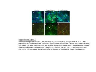
This supplementary figure (1) presents immunostaining results for HIF-1α (A-C) and HIF-2α (D-F) in skeletal muscle subjected to control (A,D), 7-day stretch (B,E), and 7-day prazosin treatment (C,F). Panels A-C feature DAPI counterstaining for nuclear visualization (blue), while panels D-F are counterstained with lectin to highlight capillaries (red). Representative images were captured at 400x magnification. Arrows indicate positive myonuclear staining for HIF proteins, and the arrowhead in Panel B highlights signaling within the pericapillary region.

Immunostaining Analysis of HIF-1α and HIF-2α in Muscles Under Stretch and Prazosin Treatment
E N D
Presentation Transcript
Control 7 day Stretch 7 day Prazosin A B C D E F Supplementary Figure 1: Immunostaining of HIF-1a (A-C) and HIF-2a (D-F) in control (A,D), 7 day stretch (B,E) or 7 day prazosin (C,F) -treated muscles. Panels A-C were counter-stained with DAPI to visualize nuclei (blue) and panels D-F were counterstained with lectin to visualize capillaries (red). Representative images of each condition were obtained at a magnification of 400x. Arrows point to positive myonuclear staining for HIF-a proteins. Arrowhead in Panel B points to signal within the pericapillary area.