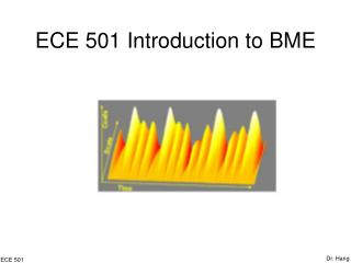
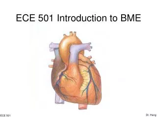
ECE 501 Introduction to BME
ECE 501 Introduction to BME. Dr. Hang. ECE 501. Part IV Bioinstrumentation Electrocardiogram. Dr. Hang. ECE 501. Introduction. Number of deaths for leading causes of death in US (2004)* Heart disease: 654,092 Cancer: 550,270 Stroke (cerebrovascular diseases): 150,147

ECE 501 Introduction to BME
E N D
Presentation Transcript
ECE 501 Introduction to BME Dr. Hang ECE501
Part IV BioinstrumentationElectrocardiogram Dr. Hang ECE 501
Introduction • Number of deaths for leading causes of death in US (2004)* • Heart disease: 654,092 • Cancer: 550,270 • Stroke (cerebrovascular diseases): 150,147 • Chronic lower respiratory diseases:123,884 • Accidents (unintentional injuries): 108,694 • Diabetes: 72,815 • Alzheimer's disease: 65,829 • Influenza/Pneumonia: 61,472 • Nephritis, nephrotic syndrome, and nephrosis: 42,762 • Septicemia: 33,464 * http://www.cdc.gov/nchs/fastats/lcod.htm Dr. Hang ECE 501
Introduction • Physiology • Instrumentation • Medicine Dr. Hang ECE 501
Physiology • What is the Heart? • The heart is a very specialized muscle that pumps • blood through the body, transporting oxygen, carbon • dioxide, nutrients and waste. • The heart is located in the middle of the chest, between • the lungs. Its bottom is tipped to the left. Dr. Hang ECE 501
Physiology • The Heart as a Pump • The heart is like two pumps: one pumping blood into the • body and one pumping blood out of the body. • The heart is about as big as two clenched fists put • together. • The heart pumps blood in beats. Dr. Hang ECE 501
Physiology • The Hard Work of the Heart Dr. Hang ECE 501
Physiology • Anatomy of the Heart Dr. Hang ECE 501
Physiology • Heart Chambers • •Atrium is a chamber that pumps blood into the heart. • Ventricle is a chamber that pumps blood out of the • heart. • The atria and the ventricles regulate blood flow by • pumping blood in and out of the heart. Dr. Hang ECE 501
Physiology • Heart Chambers Dr. Hang ECE 501
Physiology • Heart Valves • • There are four valves in the heart. • These are unidirectional valves that allow blood flow in • only one direction. • They prevent blood from flowing back to the chamber • that it has just left. Dr. Hang ECE 501
Physiology • Heart Valves Dr. Hang ECE 501
Physiology • Heart Valves • The tricuspid valve and the mitral valve are also called • A-V valves, because they separate an atrium from a • ventricle. • The pulmonary valve and the aortic valve are also called • arterial valves, because they separate a ventricle from • an artery. Dr. Hang ECE 501
Physiology • Arteries and Veins • Artery is a blood vessel that delivers blood out of the • heart. The two arteries of the heart are connected to • ventricles. • • Vein is a blood vessel that delivers blood into the heart. • The two veins of the heart are connected to atria. Dr. Hang ECE 501
Physiology • Arteries and Veins Dr. Hang ECE 501
Physiology • Pulmonary Circulation Dr. Hang ECE 501
Physiology • Pulmonary Circulation Dr. Hang ECE 501
Physiology • Pulmonary Circulation Dr. Hang ECE 501
Physiology • Systemic Circulation Dr. Hang ECE 501
Physiology • Systemic Circulation Dr. Hang ECE 501
Physiology • Systemic Circulation Dr. Hang ECE 501
Physiology • Blood Vessels Dr. Hang ECE 501
Physiology • Diffusion • The exchange of molecules between cells and blood occurs at the capillary level. • Capillaries are very small blood vessels with very thin walls. • Oxygen and nutrients diffuse from the blood into the cell and carbon dioxide and waste diffuse from the cell into the blood. Dr. Hang ECE 501
Physiology • The Cardiac Cycle: Phase I Dr. Hang ECE 501
Physiology • The Cardiac Cycle: Phase II Dr. Hang ECE 501
Physiology • The Cardiac Cycle: Phase III Dr. Hang ECE 501
Physiology • The Cardiac Cycle: Phase IV Dr. Hang ECE 501
Physiology • The Cardiac Cycle: Phase V Dr. Hang ECE 501
Physiology • The Cardiac Cycle: Phase VI Dr. Hang ECE 501
Physiology • The Cardiac Cycle: Phase VII Dr. Hang ECE 501
Physiology • Muscle Types: Skeletal Muscle • – Fast-twitching • – Voluntary control • – Gets tired • – Arms, legs etc. Dr. Hang ECE 501
Physiology • Muscle Types: Smooth Muscle • – Slow-twitching • – Involuntary control • – Does not get tired • – Stomach, bladder, blood vessels etc. Dr. Hang ECE 501
Physiology • Resting Membrane Potential Dr. Hang ECE 501
Physiology • Resting Membrane Potential • Goldman Equation Dr. Hang ECE 501
Physiology • Action Potential Once the cell is electrically stimulated (typically by an electric current from an adjacent cell), it begins a sequence of actions involving the influx and efflux of multiple cations and anions that together produce the action potential of the cell, propagating the electrical stimulation to the cells that lie adjacent to it Dr. Hang ECE 501
Physiology • Action Potential: Phase 4 Phase 4 is the resting membrane potential. This is the period that the cell remains in until it is stimulated by an external electrical stimulus (typically an adjacent cell). Dr. Hang ECE 501
Physiology • Action Potential: Phase 0 Phase 0 is the rapid depolarization phase. The slope of phase 0 is determined by the maximum rate of depolarization of the cell and is known. This phase is due to opening of the fast Na+ channels and the subsequent rapid increase in the membrane conductance to Na+ and a rapid influx of ionic current in the form of Na+ ions into the cell. Dr. Hang ECE 501
Physiology • Action Potential: Phase 1 Phase 1 of the action potential occurs with the closure of the fast Na+ channels. The transient net outward current causing the small downward deflection of the action potential is due to the movement of K+ and Cl- ions. Dr. Hang ECE 501
Physiology • Action Potential: Phase 2 This "plateau" phase of the cardiac action potential is sustained by a balance between inward movement of Ca2+ through calcium channels and outward movement of K+ through the potassium channels Dr. Hang ECE 501
Physiology • Action Potential: Phase 3 During phase 3 of the action potential, the Ca2+ channels close, while the K+ channels are still open. This ensures a net outward current, corresponding to negative change in membrane potential, This net outward, positive current (equal to loss of positive charge from the cell) causes the cell to repolarize. Dr. Hang ECE 501
Physiology • “Electrical Circuit” of the Heart Dr. Hang ECE 501
Physiology • The S-A node • The S-A Node is the most important element in the • electrical circuit of the heart. • • It starts the cardiac cycle by periodically generating • action potentials without any external stimulation. • (Therefore, it is said to be autorhythmic.) • • It is also known as the pacemaker of the heart. Dr. Hang ECE 501
Physiology • The A-V node • The atrioventricular node periodically receives action • potentials via the junctional fibers. • • The most important function of the A-V node is to • regulate the timing of the ventricular contraction by • delaying the action potentials. • • The delayed action potentials are spread over the • ventricles to cause a contraction Dr. Hang ECE 501
Physiology • The Electrical Cycle Dr. Hang ECE 501
Physiology • The Electrical Cycle Dr. Hang ECE 501
Physiology • The Electrocardiogram • The electrocardiogram (ECG) is a standardized • way to measure and display the electrical • activity of the heart. • • Physicians can diagnose problems with the • heart by analyzing its ECG and comparing it to • the ECG of a healthy heart. Dr. Hang ECE 501
Physiology • ECG Waves Dr. Hang ECE 501
Physiology • ECG Intervals Dr. Hang ECE 501