Download
1 / 1
10 likes | 184 Vues
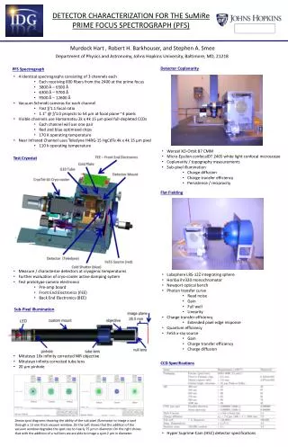
Murdock Hart , Robert H. Barkhouser , and Stephen A. Smee. Department of Physics and Astronomy, Johns Hopkins University, Baltimore, MD, 21218. Detector Coplanarity. PFS Spectrograph. 4 identical spectrographs consisting of 3 channels each

E N D
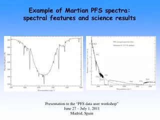
Murdock Hart , Robert H. Barkhouser, and Stephen A. Smee Department of Physics and Astronomy, Johns Hopkins University, Baltimore, MD, 21218 Detector Coplanarity PFS Spectrograph • 4 identical spectrographs consisting of 3 channels each • Each receiving 600 fibers from the 2400 at the prime focus • 3800 Å – 6500 Å • 6300 Å – 9700 Å • 9500 Å – 12600 Å • Vacuum Schmidt cameras for each channel • Fast ƒ/1.1 focal ratio • 1.1” @ ƒ/1.0 projects to 54 μm at focal plane ~4 pixels • Visible channels use Hamamatsu 2k x 4k 15 μm pixel full-depleted CCDs • Each channel will use one pair • Red and blue optimized chips • 170 K operating temperature • Near Infrared Channel uses Teledyne H4RG-15 HgCdTe 4k x 4k 15 μm pixel • 110 k operating temperature DETECTOR CHARACTERIZATION FOR THE SuMiRe PRIME FOCUS SPECTROGRAPH (PFS) • Wenzel XO-Orbit 87 CMM • Micro-Epsilon confocalDT 2405 white light confocal microscope • Coplanarity / topography measurements • Sub-pixel illumination • Charge diffusion • Charge transfer efficiency • Persistence / reciprocity Test Cryostat Flat Fielding • Measure / characterize detectors at cryogenic temperatures • Further evaluation of cryo-cooler active damping system • Test prototype camera electronics • Pre-amp board • Front End Electronics (FEE) • Back End Electronics (BEE) • Labsphere LRS-12Z integrating sphere • Horiba ihr320 monochromator • Newport optical bench • Photon transfer curve • Read noise • Gain • Full well • Linearity • Charge transfer efficiency • Extended pixel edge response • Quantum efficiency • Fe55 x-ray source • Gain • Charge transfer efficiency • Charge diffusion Sub-Pixel Illumination • Mitutoyo 10x infinity corrected NIR objective • Mitutoyo infinity corrected tube lens • 20 μm pinhole CCD Specifications Zemax spot diagrams showing the ability of the sub pixel illuminator to image a spot through a 13 mm thick vacuum window. On the Left shows that the addition of the vacuum window degrades the spot size to nearly 75 μm in diameter. On the right shows that with the addition of a null lens we are able to image a spot 2 μm in diameter. • Hyper Suprime-Cam (HSC) detector specifications