Download
1 / 42
440 likes | 782 Vues
Chronic Obstructive Pulmonary Disease. By: Sara-Louise Walsh, SPT. Objectives:. After this in-service presentation: Viewers will be able to accurately describe how COPD damages the body when prompted.

E N D
Chronic Obstructive Pulmonary Disease By: Sara-Louise Walsh, SPT
Objectives: • After this in-service presentation: • Viewers will be able to accurately describe how COPD damages the body when prompted. • Viewers will be able to correctly name 3 physical therapy interventions that help patients who have COPD when prompted.
What is COPD? • Progressive • Irreversible Lung Disease • Characterized by chronic bronchial outflow obstruction
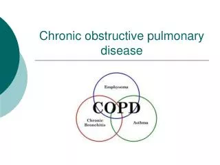
What is COPD? A category of lung diseases. • Includes: • Emphysema • Chronic Bronchitis • Asthma • Bronchiectasis
Terminology • COPD often begins as emphysema or chronic bronchitis. • These two diseases are similar in progression. • If a patient is diagnosed with a lung disease after years of smoking it may be difficult to identify which disease the pt has. • A patient who has one disease is likely to have another disease under the category of COPD as well.
Common Pathology • Decreased ability of body to move air into and out of the lungs • Causes decreased oxygen intake and carbon dioxide output. • Body is unable to use oxygen to produce energy.
*Trivia* • Question: • What is the most common cause of COPD?
Answer: • Cigarette Smoking
Emphysema • Initial Lesion: • Destruction of alveolar walls • Alveoli merge • Large air spaces form (bulla or bleb) • 90% caused by smoking
Results • Inflames lungs • Inhibit alpha-1 antitrypsin- • neutralize digestive enzymes produced by inflammatory cells • prevent over activity and digestion of normal tissue.
How Common is Emphysema? • Common • Present in half of all autopsy reports. • most had no symptoms
Visual of a Typical Emphysema Patient • Pink Puffer • Thin • Smoker • Short of breath • Barrel shaped chest • Breaths thru pursed lips • Causes pink cheeks • Wheezing • Coughing
Cont. • Over muscular neck muscles • Pt bends forward at the waste to ease breathing • Infection • Sputum production
Clinical Description of a Typical Emphysema Patient • Short of breath= first symptom • Dramatic wt loss • body sheds muscle the lungs can not support
*Trivia* • What might a typical emphysema patient look like?
Answer • Thin • Smoker • Short of breath • Barrel shaped chest • Breaths thru pursed lips • Wheezing • Coughing • Over muscular neck muscles • Pt bends forward at the waste to ease breathing • Infection • Sputum production
Chronic bronchitisDefinition: • Chronic cough that produces sputum for 3 consecutive months two years in a row. • Primary Cause: cigarette smoking
Chronic Bronchitis:Results: • Initial Lesion: Bronchi • Smoke irritates bronchi and stimulates chronic inflammation and bronchial mucous secretion.
Chronic BronchitisConsequences: • Simple chronic bronchitis • Productive cough • Do not have airway obstruction • Chronic asthmatic bronchitis • Some periods of asthma-like wheezing • Obstructive chronic bronchitis • Consistent wheezing and obstruction
Relationship Between Emphysema and Chronic Bronchitis • Most patients with chronic bronchitis also have emphysema
Visual of a Typical Patient who has Chronic Bronchitis • Blue Bloater • Hypoxic • Large Mid-section • No weight loss
Clinical Description • Airflow obstruction • Wheezing • Coughing • Sputum production • Infection • Acidotic
Note: • Most patients visually fall somewhere between the “pink puffer” and “blue bloater” examples.
*Trivia* • Name two lung diseases a smoking patient is likely to have simultaneously.
Answer: Emphysema & Chronic Bronchitis
Bronchiectasis • Marked permanent dilation of small bronchi • Destruction of smooth m and elastic supporting tissue of the airway • Both obstruction and infection are required-either may come first.
Bronchiectasis:Cause: • Usually secondary to something else • Immunodeficiency states-encourage infection • Chronic infection such as tuberculosis- damage bronchial walls • Retained bronchial foreign body-mucous retention and infection • Cystic fibrosis-prod thick mucous that obstructs bronchi
Bronchiectasis:Results: • Obstruction can cause mucous retention-breeding ground for infection • Lower lobes typically involved, especially in airways that are close to vertical • Dilated flaccid, pus filled tube
Bronchiectasis:Clinical Description of a Typical Patient: • Chronic Infection • Persistent Cough • Production of a large amount of yellow, fowl-smelling sputum.
*Trivia* • Name one component of the clinical description of a patient who has bronchiectasis.
Answer: • Chronic Infection • Persistent Cough • Sputum Production
Results of COPD • Respiratory muscle fatigue • Cardiovascular deconditioning • Poor Posture • Rounded shoulders • Forward head • Anterior accessory muscles compensate for breathing. • Death from COPD is usually associated with a combination of hypoxia, acidosis, and right-sided heart failure. • Pneumonia is often the terminal event
Special Considerations • Bronchodilators may cause tachycardia or muscle weakness. • All pts with COPD are at risk for chronic hypoxia (low oxy) • Can lead to pulmonary vasospasm • Pulmonary hypertension • Right-sided heart failure
What kind of physical therapists see patients who have COPD? • Hospital Setting • In-patient • Out-patient • Private Practice • Nursing Home • Home Care • Researchers
Specific Physical Therapy Goals: • Improve Quality of Life = Improve Ability to Breathe • Improve Breathing Pattern • Increase exercise tolerance • Improve Posture • Promote Relaxation of Accessory Muscles of Inspiration
Interventions: Improve Breathing Pattern • Diaphragmatic Breathing • Pursed Lip Breathing • Encourage Cessation of Smoking Habits • 20 minutes after quitting: heart rate and blood pressure drop • 12 hours after quitting: carbon monoxide levels normalize • 2-3 months after quitting: lung function improves and circulation increases • 1 year after quitting: risk of coronary artery disease is cut in half
Interventions: Increase Exercise Tolerance • Graded Endurance and Conditioning Exercises • Aerobic training • 30 minutes/day • 11-13 on RPE • Balance Training • Strength Training for Lower Extremities
Interventions: Improve Posture • Strength training • Low resistance • High reps • 2-3 Days/Week • Flexibility training • Position for Relaxation
*Trivia* • Name three physical therapy interventions for patients who have COPD.
Answer • Improve Breathing Pattern • Increase Exercise Tolerance • Improve Posture
Interesting Research • One-legged exercise training • Use of supplemental oxygen
Works Cited • Como D. Mosby’s Dictionary 8th edition. St. Louis, MO. Elselvier. 2009. 379. • McConnell, TH. The Nature of Disease. Baltimore, MD. Lippincott Williams & Wilkins. 2007. 332-335. • Gardenhire D. Adrenergic bronchodilators and side effects: what do you look for? Journal for respiratory care and sleep medicine. 2008. March • Kisner C, Colby LA. Therapeutic Exercise. 5th edition Philadelphia, PA. Davis. 2007. 864-877. • Dolmage TE, Goldstein RS. Effects of one-legged exercise training in patients with copd. Chest 2008; 133 (2) 370-376 • Jolly EC, Di Boscio V, Aguirre L, Luna CM, Berensztein S, Gene RJ. Effect of supplemental oxygen during activity in patients with advanced COPD without severe resting hypoxemia. Chest. 2001; 120 (2): 437-443.