Download
1 / 83
830 likes | 1.02k Vues
COST B27 ENOC Joint WGs Meeting Swansea UK, 16-18 September 2006. SLEEP , AUTONOMIC CONTROL AND PSYCHOEMOTIONAL STATUS. Giedrius Varoneckas. Institute of Psychophysiology and Rehabilitation c/o Kaunas University of Medicine Vyd ū no Str. 4, Palanga LT-00135, Lithuania

E N D
COST B27 ENOC Joint WGs Meeting Swansea UK, 16-18 September 2006 SLEEP, AUTONOMIC CONTROL AND PSYCHOEMOTIONAL STATUS Giedrius Varoneckas Institute of Psychophysiology and Rehabilitation c/o Kaunas University of Medicine Vydūno Str. 4, Palanga LT-00135, Lithuania E-mail: giedvar@ktl.mii.lt
The goal of this presentation Demonstration ofadiagnostic value of HR variability analysisas well asrelationshipbetweendepression/anxiety, sleep quality, cardiovascular function andautonomic control From other hand, HR variability biofeedback training is powerful tool for treatment of this various disorders
Background: What is Biofeedback? • Biofeedback is a treatment technique in which people are trained to improve their health by using signals from their own bodies • “Biofeedback" was coined in the late 1960s to describe laboratory procedures then being used to train experimental research subjects to alter brain activity, blood pressure, heart rate, and other bodily functions that normally are not controlled voluntarily • Psychologists use it to help tense and anxious clients learn to relax Bette Runck. DHHS Publication No (ADM) 83-1273
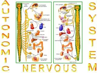
Background: Biofeedback & Autonomic Control • By providing access to physiological information about which the user is generally unaware, biofeedback allows users to gain control over physical processes previously considered automatic • Interaction between central nervous system and autonomic control plays role in “biofeedback” process • The biofeedback response is related to the baseline level of autonomic control Jacenko M. Wikimedia Commons
Background: Autonomic Control Modifications • During wake-sleep cycle as a reflection of brain functions • In depression/anxiety • In somatic disorders (coronary artery disease) • In sleep restriction
Autonomic HR control goes through three main mechanisms • balancebetween of sympathetic-parasympathetic branches of autonomic nervous system (HR frequency and oscillatory structure) • tonic control(HR variability), depending of P/S interaction • reflex control(mainly baroreflex) level might be drawn from assessment of baroreflex sensitivity and/or HR maximal response to AOT
Wakefulness (active) 50 mV 1 sec Wakefulness (passive) Stage 1 Theta waves Stage 2 Sleep spindles Stage 4 “Saw teeths” REM Sleep Sleep Electroencephalography K-complex
Normal Sleep HistogramSequences of States and Stages of Sleep on a Typical Night Identification and Staging of Adult Human Sleep. L.Shigley. Sleep Academic Award
Heart Rate and Heart Rate Variability during Sleep Zemaityte D. et al. Psychophysiology, 1984, 21(3), 279-289
Methods of obtaining the HRV parameters may by divided into following groups: Time domain methods Spectral domain methods Non-linear methods Mathematic modeling methods
HR analysis using power spectrum Three main oscillatory components: very low frequency component (VLFC) low frequency component (LFC) high frequency component (HFC) in absolute (ms) and relative (percent) values for evaluation: humoral,sympathetic-parasympathetic and parasympathetic control, correspondingly
Heartrate analysisusing Poincare plot RRr, difference on plot diagonal between minimal (RRmin) and maximal (RRmax) RR values RRt, maximal HR variability as maximal width-difference between of two points at parallel tangential lines determining plot RRmin, maximalHR frequency RRmax, HR frequency at its minimal level RRrt RRr RRmin RRmax P, square of the plot, representing overall HR variability
MethodsNonlinear analysis of continuous ECG during sleep: II. Dynamical measures Fell J. et al. Biol. Cybern. 82, 485-91 (2000) The correlation dimension serves as an estimator of the number of degrees of freedom in a system, this is, the number of variables required to generate the observed dynamics D2 - as a measure of the complexity of a time series (Grassberger & Proccacia 1983) ECG dynamics was considered to be composed of two aspects: (i) the inter-beat or RR variability; (ii) the PQRST complex Tab. Contrasts between sleep stages for the nonlinear ECG measures D2, L1, K2 and average first return time (p = 0.05) • An increase in dominant chaoticityduring REM sleep with regard to time-continuous nonlinear analysis is comparable to an increased heart rate variability • The reduction in the correlation dimension (D2) may be interpreted as an expression of the withdrawal of respiratoryinfluences during REM sleep
Methods: Detrended fluctuation analysis Comparison of detrended fluctuation analysis and spectral analysis for HRV in sleep and sleep apnea: 14 healthy subjects, 33 pts with moderate, and 31 pts with severe sleep apnea VLF, LF, HF, and LF/HF confirmed increasing parasympathetic activity from wakefulness and REM over light sleep to deep sleep, which is reduced in patients with sleep apnea Changes in HRV are better quantified by scaling analysis than by spectral analysis Penzel T. et al. IEEE Trans Biomed Eng. 2003;50(10):1143-51.
Methods: Correlation dimension (D2)Application of chaos theory in analyzing the HR in healthy subjects during sleep stages The correlation between the changes in D2 during different sleep stages and the level of autonomic HRcontrol was demonstrated The chaotic element of HR, expressed numerically by D2 depends on the baseline level autonomic HR control Eidukaitis A. et al. Human Physiology, 2004, 30, 5, 551-5.
The heart rate during synchronized sleep after different steps of heart denervationExperiment from unrestrained cats with chronically implanted electrodes Intact Baust W. & Bohnert B.The Regulation of Heart During SleepExp. Brain Res. 7, 169-180 (1969) BilateralStellatectomy Neurological Clinic, University of Düsseldorf Germany BilateralVagotomy Combined Stellatectomy& Vagotomy
Changes in heart rate during shift from synchronize to desynchronized sleepExperiment from unrestrained cats with chronically implanted electrodes Intact BilateralVagotomy Combined Stellatectomy& Vagotomy Non-REM Sleep → REM Sleep Baust W. & BohnertB. Exp. Brain Res. 7, 169-180 (1969)
Sympathetic and parasympathetic control study Egberg & Katona modification of the model suggested by Rosenblueth & Simeone (Am J Physiol, 1934, 110, 42-55) Egberg JR and Katona PG,Am. J Physiology, 1980, 238, H829-H835.
Sympathetic (S) and parasympathetic (P) multipliers’values as functions of sleep S P 1st night – adaptation 2nd night – intact 3rd night - atropine 0.025 mg/kg 4th night - propranolol retard 160 mg 5th night – atropine plus propranolol Zemaityte D. et al. Psychophysiology, 1984, 21(3), 279-289
Poincare plots of RR intervals in healthy subject under different conditions of autonomic HR control Propanolol &Atropine Baseline Propranolol Atropine Wakefulness Stage 2 Stage 3 REM Sleep
Heart rate, stroke volume, and cardiacoutputas a functions of sleep stages RR, s SV, ml CO, l/min RR, % SV, % CO, % W 1 2 3 4 REM W 1 2 3 4 REM W 1 2 3 4 REM Healthy Ss Zemaityte D. et al. Psychophysiology, 1984, 21(3), 279-289&290-298 CAD Pts Varoneckas G. Fiziologija Cheloveka, 1994, 20, 1, 76-83.
Heart Rate Sleep Patternin Healthy Subject W 1 2 3 4 REM Zemaityte D. et al. Psychophysiology, 1984, 21(3), 279-289
Poincare plots of RR intervals during individual sleep stages W before sleep REM Sleep Overall night Stages 1-2 Stages 3-4 Healthy Subject Typical HRSP CAD Patients Reduced HRSP Zemaityte D. et al. Biomedicine, 2001, 1, 1, 34-44
Dynamic heart rate variability: a tool for exploring sympathovagal balance continuously during sleep in men Hélène Otzenberger, Claude Gronfier, Chantal Simon, Anne Charloux, Jean Ehrhart, François Piquard, and Gabrielle Brandenberger Laboratoire des Régulations Physiologiques et des Rythmes Biologiques chez l'Homme, Institut de Physiologie, 67085 Strasbourg Cedex, France Am J Physiol Heart Circ Physiol 1998, 275: H946-H950 Fig. 1. Examples of 5-min Poincaré plots with regard to power spectra of R-R intervals, according to sleep states: intrasleep awaking (W), stage 2 (St2), slow-wave sleep (SWS), and rapid eye movement sleep (REM). rRR, Interbeat autocorrelation coefficient of R-R intervals; R-Rn and R-Rn+1, successive R-R intervals; LF/HF, ratio of low- to high-frequency power.
Heart Rate Sleep Pattern Reduced Typical Zemaityte D. et al. Psychophysiology, 1984, 21(3), 279-289Zemaityte D. et al. Psychophysiology, 1984, 21(3), 290-298
Changes of HR power spectrum components impact during shifts of sleep stages Healthy Ss- Typical HRSP CAD pts – Typical HRSP CAD pts – Reduced HRSP W Stage 1 Stage 2 Stage 3 Stage 4 REM VLFC, % LFC, % HFC, % Zemaityte et al. Int J Psychophysiology, 1986, 4, 129-141
Effects of Aging and Cardiac Denervation on Heart Rate Variability During Sleep RR intervaldid not differ between young and older subjects during awake periods but was larger in the young subjects than in the older subjects during both non-REM and REM The total RR variability was higher in the young subjects than in the older volunteers during awake, non-REM sleep, and REM sleep Figure 1. RR during awake periods, non-REM, and REM sleep in normal young (open bars) and older (solid bars) subjects. Reduction in RR with aging is particularly evident during non-REM and REM sleep. +P<0.05, •P<0.01 vs old; ••P<0.01, XXP<0.001 vs awake. Crasset V. et al. Circulation. 2001;103:84
Effects of Aging and Cardiac Denervation on Heart Rate Variability During Sleep • In conclusion, study reveals • that the normal, age-related disappearance of deep sleep impairs the nocturnal increase in cardiac vagal activity and • that nonautonomic mechanisms slightly increase HF oscillations in RR during non-REM sleep in humans. Figure 2. Effects of aging on RR variability during non-REM and REM sleep. HF oscillations in RR became more predominant than LF oscillations in RR during non-REM sleep in normal young subjects (open bars). These changes were lost with aging (solid bars). +P<0.05 vs old; ++P<0.05 vs awake. Crasset V. et al. Circulation. 2001;103:84
Vanoli E. et al. Heart Rate Variability in Specific Sleep Stages: A Comparison of Healthy Subjects and Patients After Myocardial Infarction. Circulation,1995; 91: 1918-1922. Spectral analyses of heart rate variability during non-rapid eye movement sleep from one normal individual and in a patient with a recent myocardial infarction Sleep contains information that is highly relevant to the identification of autonomic derangements associated with a higher risk for lethal events after MI. Expected surge in cardiac vagal activity associated with non-REM sleep is completely lost after MI. The higher risk for ischemic events and the unopposed sympathetic activity evident during REM sleep creates a condition in which lethal arrhythmic events are more likely to occur and provide new information to the understanding of sudden death at night.
Poincare plots and power spectra of all-night HR recording Sportsman Healthy subject CAD patient Kesaite R..et al.Information Technology and Control, 2001, 2, 20-28
The restorative function of sleep towards the cardiovascular system in trained sportsmen and non-trained healthy Ss Trained sportsmen Non-trained healthy Ss Varoneckas G. Fiziologija Cheloveka, 1994, 20, 1, 76-83.
HRvariability inwell-trained sportsman and non-trained subject well-trained sportsman non-trained subject
The restorative function of sleep towards the cardiovascular system in healthy Ss and CAD pts Healthy Ss CAD pts Varoneckas G. Fiziologija Cheloveka, 1994, 20, 1, 76-83.
Sleep AOT evening-time AOT morning-time HR variability during sleep and AOT in healthy subject and CAD patient Healthy subject CAD pt
HRvariabilityin CAD patient with andwithout HR restoration with HR restoration inability to restore
Total sleep time Healthy Subjects Pts with HR restoration CAD Patients Pts showing inability to restore HR min min *
Sleep structure in CAD patients with Restoration of HR control Inability to restore HR min * p<.05 * * *
HR variability in healthy subject and CAD pts surviving and died during 2-yr follow-up Healthy Survived Died Poincare maps from successive RR intervals during night sleep and exercise Zemaityte D. et al. Biomedicine, 2002, 2, 1, 2-14.
Poincare maps from successive RR intervals during sleep and exercisein cardiac pts with dysrrhythmias SPT Healthy Ss VTT PAF Zemaityte D. et al. Biomedicine, 2002, 2, 1, 2-14
Prognostic value of ventricular arrhythmias and heart rate variability in patients with unstable angina Lanza GA et al. Heart. 2005 Dec 30 • 17 cardiological centres in Italy • 543 patients with UA and EF>40% • Holter ECG within 24 h of hospital admission • Adjusted for clinical (age, gender, cardiac risk factors, history of previous MI) and laboratory (troponin I, C-reactive protein, transient myocardial ischemia on HM) variables 3 HRV variables (standard deviation of RR intervals index, low frequency [LF] amplitude and low to high frequency ratio [LF/HF]) were associated with in-hospital death, and bottom quartile values of most HRV variables predicted 6-month fatal events
Prognostic value of ventricular arrhythmias and heart rate variability in patients with unstable angina Lanza GA et al. Heart. 2005 Dec 30 Table 2. Variables significantly predictive of in-hospital death at univariable analysis. Complex ventricular arrhythmias are not included as all patients who died showed these forms of arrhythmias on Holter monitoring. • 17 cardiological centres in Italy • 543 patients with UA and EF>40% • Holter ECG within 24 h of hospital admission • Adjusted for clinical (age, gender, cardiac risk factors, history of previous MI) and laboratory (troponin I, C-reactive protein, transient myocardial ischemia on HM) variables Complex VA and frequent PVBs were strongly predictive of death in hospital and at follow-up In UA patients with preserved myocardial function, both VA and HRV are independent predictors of in-hospital and medium-term mortality
HR variability in evaluation of autonomic control and cardiovascular status in patients after cardiac surgery Before CABG surgery After 1 mos After 6 mos Poincare maps from successive RR intervals during sleep and exercisein cardiac patient Zemaityte D. et al. Biomedicine, 2002, 2, 1, 2-14
Impairment of cardiovascular autonomic control in patients early after cardiac surgery 25 male patients underwentCABG surgerynormal values were obtained from healthy volunteers Baroreflex function, HR variability and blood pressure variability in patients early after coronary surgery Obviously, there is a vagal suppression 20 h after surgery, while the sympathetic tonus works in a normal range This unbalanced interaction of the autonomous systems is similar to findings in patients after myocardial infarction Bauernschmitt R. et al. Eur J Cardiothorac Surg. 2004;25(3):320-6.
Modification of HR variability due to medication by ACEI Before After 1 mo After 12 mos Poincare maps from successive RR intervals during night sleep and exercise
Changes of total HR variability during night sleep due to ACEI treatment p < .05 during sleep RR, ms p < .05 after sleep p < .05 p < .05 p < .05 sRR, ms before sleep
Gujjar AR et al. Heart rate variability and outcome in acute severe stroke: role of power spectral analysis. Neurocrit Care. 2004;1(3):347-53. • 25 patients: 11 died, 10 - poor, and 4 - good outcome • 6 comatose patients (Glasgow Coma score<9 at admission)16 had focal weakness all had abnormal brain computed tomography • HRV parameters from continuous 1-hour record • Low-frequency (LF) and very low-frequency (VLF) spectral power correlated with mortality HRV measurements are independent predictorsof outcome in acute severe stroke
Heart rate, systolic and diastolic blood pressure and baroreflex sensitivity during sleep
RR ms mmHg SystolicBP Diastolic BP BRS ms/mmHg Baroreflex sensitivity, heart rate and blood pressure during sleep Healthy Ss n=16 CAD pts n=48
Baroreflex sensitivity during different sleep stages in CAD patients with sleep apnea BRS ms/mmHg AHI5 n=59 5>AHI15 n=29 AHI>15 n=31 The groups were matched according age, body mass index and cardiac pathology
RR ms Systolic BP mmHg Diastolic BP ms/mmHg BRS Baroreflex sensitivity, heart rate and blood pressure during sleep in CAD patients Without VPBs (57 pts) With VPBs (62 pts) • The groups were matched according age, body mass index and cardiac pathology