Download
1 / 42
430 likes | 1.1k Vues
Nutrition, Digestion, Absorption, and Nitrogen Excretion Adaptations for Feeding Heterotrophic organisms can be classified by how they acquire their nutrition. Decomposers , mostly protists and fungi, absorb nutrients from dead organic matter.

E N D
Adaptations for Feeding • Heterotrophic organisms can be classified by how they acquire their nutrition. • Decomposers, mostly protists and fungi, absorb nutrients from dead organic matter. • Detritivores, such as earthworms and crabs, actively feed on dead organic material. • Predators are animals that feed on living organisms: • Herbivores prey on plants. • Carnivores prey an animals. • Omnivores prey on both.
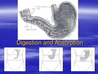
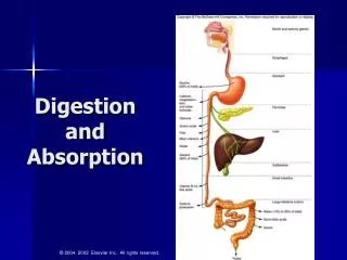
Digestion • Food is taken into a body cavity that is continuous with the outside environment, where it is acted on by enzymes secreted by the animals. • These enzymes break the food down into nutrient molecules that are absorbed by the cells lining the cavity. • Most animals have a tubular gut with a mouth that takes in food and an anus for waste excretion.
Digestion • The anterior end of the gut consists of the mouth and the buccal cavity (mouth cavity). • Food is usually broken up here into smaller fragments by structures such as teeth. • Stomachs are storage chambers that enable animals to ingest large amounts of food and digest it at leisure. • The next section of the gut is called the midgut, or intestine. Most materials are digested and absorbed here. • Specialized glands secrete some digestive enzymes into the intestine, and cells in the gut wall itself secrete other digestive enzymes.
Digestion • The hindgut recovers water and ions and stores undigested wastes (feces). • A muscular rectum near the anus assists in the expulsion of feces (defecation). • Many species have colonies of endosymbiotic bacteria within their hindguts. • These bacteria obtain nutrients from the food passing through the host’s gut and contribute to the digestive processes of the host.
Digestion • The parts of the gut that absorb nutrients have large surface areas to maximize nutrient absorption. • Vertebrates have a gut wall that is richly folded, with individual folds bearing fingerlike projections called villi, which in turn have projections called microvilli.
Figure 50.9 Greater Intestinal Surface Area Means More Nutrient Absorption (Part 3)
Structure and Function of the Vertebrate Gut • The vertebrate gut has four layers of tissue throughout its length. • The cavity of the gut is called the lumen. • Starting from the lumen, the first layer of tissue is the mucosa. • Cells of the mucosa have secretory and absorptive functions. • Just outside the mucosa is the second layer of cells, the submucosa, which contains blood and lymph vessels that carry absorbed nutrients to the rest of the body. • There are two layers of smooth muscle cells external to the submucosa.
Structure and Function of the Vertebrate Gut • When food enters the mouth it is chewed and mixed with the secretions of salivary glands. • When the food makes contact with the back of the mouth, the reflexive action of swallowing is initiated. • Swallowing involves muscles propelling food through the pharynx (where the mouth cavity and the nasal passages join) and into the esophagus (the food tube).
Figure 50.12 Swallowing and Peristalsis (Part 1) Bolus – a rounded, semisolid mass of food that is either being swallowed or passing through the digestive tract.
Structure and Function of the Vertebrate Gut • Carbohydrate digestion begins in the mouth, where amylase is secreted with saliva and mixed with the food as it is chewed. • Secretions of the stomach kill microorganisms that are taken in with food and begin the digestion of proteins. • An endopeptidase called pepsin is the major enzyme produced by the stomach. • Initially, pepsin is secreted by cells in the gastric glands in its inactive form called pepsinogen.
Structure and Function of the Vertebrate Gut • Hydrochloric acid (HCl) maintains a pH of 1 – 3 in the stomach fluid, which activates the conversion of pepsinogen to pepsin. • Mucus secreted by the stomach mucosa coats the walls of the stomach and protects them from being eroded and digested by HCl and pepsin. • When walls of the stomach are exposed directly to HCl and pepsin, an ulcer can result.
Structure and Function of the Vertebrate Gut • The muscles in the walls of the stomach contract to churn its contents and mix them with the stomach secretions. • Peristaltic contractions of the stomach push the digested food toward the bottom end of the stomach and into the beginning of the intestine through the pyloric sphincter. • When this partially digested food leaves the stomach, then it is called chyme.
Structure and Function of the Vertebrate Gut • In the small intestine, the digestion of carbohydrates and proteins continues, and the digestion of fats and the absorption of nutrients begins. • The small intestine has three sections: The duodenum is the initial section and is the site of most digestion. • The jejunum and the ileum carry out 90 percent of the absorption of nutrients.
Structure and Function of the Vertebrate Gut • The liver and the pancreas provide many of the specialized enzymes required for digestion. • The liver produces bile, which aids in fat digestion. • Bile is secreted from the liver and flows through a branch of the hepatic duct to the gallbladder, where it stored until it is needed. • When fat enters the duodenum, bile is squeezed into the common bile duct, where it flows into the duodenum.
Structure and Function of the Vertebrate Gut • Bile is an emulsifier – a substance that prevents oil droplets from aggregating. • Bile emulsifies fats and greatly increases the surface area of the fats that are exposed to lipases. • The small fat particles surrounded by bile molecules are called micelles.
Structure and Function of the Vertebrate Gut • The pancreas is a large gland that lies just beneath the stomach and functions as both an endocrine and exocrine gland. • The exocrine tissues of the pancreas produce a number of digestive enzymes, released as zymogens, such as trypsinogen. • The pancreas also produces a secretion rich in bicarbonate ions, which neutralize the pH of the chyme from the stomach. • This process is essential because intestinal enzymes function best at a neutral or slightly alkaline pH
Structure and Function of the Vertebrate Gut • The mechanisms by which cells lining the intestine absorb nutrient molecules are diverse and not completely understood. • Carrier proteins actively transport many inorganic ions into cells. • Carrier proteins also transport amino acids, glucose, and galactose, in conjunction with diffusion of sodium ions. • The process of fat absorption does not involve carrier proteins. • Lipases break fats down into diglycerides, monoglycerides, and fatty acids, which are able pass through the plasma membrane of microvilli.
Structure and Function of the Vertebrate Gut • Peristalsis pushes the contents of the small intestine into the large intestine, or colon. • The colon absorbs water and ions, producing semisolid feces from indigestible material. • Too much water absorption results in constipation and too little water absorption results in diarrhea. • Large populations of bacteria live in the colon, including Escherichia coli, which synthesizes vitamin K and biotin that are absorbed across the wall of the colon. • Prolonged intake of antibiotics can lead to vitamin deficiency because the antibiotics kill the normal intestinal bacteria.
Structure and Function of the Vertebrate Gut • Intestinal bacteria produce gases such as methane and hydrogen sulfide as by-products of anaerobic metabolism. • A large percentage of the material in feces consists of cell walls of dead bacteria.
Excreting Nitrogenous Wastes • Fats and carbohydrates break down into water and CO2, which are easily eliminated. • Proteins and nucleic acids contain nitrogen. Breakdown of these produces nitrogenous waste. • Ammonia is the most common nitrogenous waste product and is highly toxic. • It must be quickly eliminated or converted into less toxic molecules such as urea and uric acid.
Figure 51.3 Waste Products of Metabolism ex. fishes ex. mammals, amphibians Most species produce more than one nitrogenous waste. Different developmental stages may have different forms of nitrogen excretion. ex. insects, reptiles, birds
Vertebrate Excretory Systems • The kidney is the major excretory organ of vertebrates. • The functional unit of the vertebrate kidney is the nephron. • Each human kidney has about a million nephrons.
Vertebrate Excretory Systems • Nephrons have three main parts: • The glomerulus is a ball of capillaries that filters plasma. • The renal tubules receive and modify filtrate. • Peritubular capillaries carry substances to and from the renal tubules.
Vertebrate Excretory Systems • The two capillary beds of the nephron—the glomerulus and the peritubular capillaries—lie in series between the arteriole and the venule. • An afferent arteriole supplies blood under pressure to the glomerulus; the blood leaves through an efferent arteriole. • The renal tubule begins with Bowman’s capsule which encloses the glomerulus. • Cells of the capsule that come into direct contact with the glomerular capillaries are called podocytes. They have fine projections that wrap around and cover the capillaries.
Vertebrate Excretory Systems • The glomerulus filters the blood to produce a fluid that lacks cells and large molecules (renal filtrate). • The walls of the capillaries, the basal lamina of the capillary endothelium, and the podocytes of Bowman’s capsule all participate in filtration. • The force that drives filtration in the glomerulus is the pressure of the arterial blood.
Vertebrate Excretory Systems • The composition of fluid entering the nephron is similar to that of blood plasma, except it lacks the plasma proteins. • As the fluid moves down the renal tubule, it is concentrated and altered to form urine. • The cells of the tubule control the composition of the urine by actively secreting and resorbing specific molecules.
The Mammalian Excretory System • Humans have two kidneys, which filter blood, process the filtrate into urine, and release the urine into a duct called the ureter. • The ureter of each kidney leads to the urinary bladder, where urine is stored until it is excreted through the urethra. • Two sphincter muscles surrounding the base of the urethra control the timing of urination. • One is smooth muscle, controlled by the autonomic nervous system. When the bladder is full, a spinal reflex relaxes this sphincter. • The other sphincter is skeletal muscle and is under voluntary control.
The Mammalian Excretory System • The ureter, renal artery, and renal vein enter the kidney on its concave side. • Subunits of the kidney called renal pyramids include all the functional units. • The renal pyramids make up the medulla of the kidney and are surrounded by tissue called the cortex. • Each kidney has about a million nephrons, which are its basic functional units.
The Mammalian Excretory System • The first section of a renal tubule (closest to the glomerulus) is called the proximal convoluted tubule. These lie in the cortex. • The renal tubule then dives into the medulla and makes a loop called the loop of Henle. • The tubule of the loop of Henle makes a hairpin turn within the medulla and continues back up to the cortex. • When it reaches the cortex again it is called the distal convoluted tubule. • Distal convoluted tubules join to form collecting ducts.
The Mammalian Excretory System • The organization of blood vessels in the kidney parallels the organization of the nephrons. • Arterioles branch from the renal artery and travel into the cortex. • An afferent arteriole carries blood to each glomerulus. The efferent arteriole also gives rise to the peritubular capillaries. • Most peritubular capillaries are in the cortex, but a few run down into the medulla, parallel to the loop of Henle and the collecting ducts. • In the medulla region, these capillaries form the vasa recta.
The Mammalian Excretory System • The proximal convoluted tubule has specialized cuboidal cells with thousands of microvilli which greatly increase the surface area for resorption of ions and molecules. • The cells have many mitochondria to produce the ATP needed to operate the transport systems. • They actively transport Na+ and other solutes, such as glucose and amino acids, out of the tubule lumen, and water follows by osmosis. • Almost all glucose and amino acid molecules are resorbed. • The peritubular capillaries take up the water and solutes.
The Mammalian Excretory System • Humans can produce urine that is four times more concentrated than their blood. Some mammals produce urine even more concentrated. • The loop of Henle functions as a countercurrent multiplier system to increase the solute potential of the surrounding tissue fluid. • The tubule fluid in the descending limb of the loop flows in the opposite direction from that of the ascending limb. • The system creates a solute concentration gradient in the renal medulla.
The Mammalian Excretory System • When the fluid enters the collecting duct, it is the same concentration as blood plasma, but the composition is different. • The major solute in the duct is now urea. As fluid flows down the collecting duct, it loses water osmotically. • Some urea also leaks out of the collecting duct to the surrounding tissue, which increases the osmotic potential of the tissue. • The urea diffuses back to the loop of Henle. The recycling of urea contributes to the ability to concentrate urine.