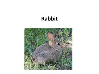
Rabbit Diseases
Rabbit Diseases. Bacterial and Mycotic Diseases.


Rabbit Diseases
E N D
Presentation Transcript
At the conclusion of this program, you should be able to state the significance and etiologic agents of the following diseases in rabbits maintained for research, testing, or teaching: Pasteurellosis, entero-toxemia, mucoid enteropathy, treponematosis, mastitis, Tyzzer's Disease, and superficial mycosis.
Most common disease of domestic rabbits Etiology: Pasteurella multocida It is a bacterial infection that gains access to the nasal cavity, then migrates to almost any organ in the body. It can migrate via the eustachian tube to the ear, via the nasolacrimal duct to the conjunctiva, or by way of the trachea to the lungs. It can be carried by the blood stream, to cause abscesses and lesions in internal organs, such as heart, lungs, and reproductive systems. It can also gain access through skin wounds to cause subcutaneous abscesses. Pasteurellosis – “Snuffles”
The classic clinical manifestation of pasteurellosis is a mucopurulent nasal discharge that can be accompanied by sneezing, coughing, and snuffling sounds. These signs account for the common name of "snuffles" that is given to the condition. Clinical Signs:
Exudate on forepaws The nasal exudate may be wiped onto the medial aspects of the forepaws Clinical signs cont…
The signs of pasteurellosis will be related to impairment of the organs that are involved. The conjunctiva may be red and swollen with serous to purulent exudate that may mat on the fur. Signs -- red eye
Torticollis, or head tilt, may be seen as the result of middle or inner ear infection, or subcutaneous abscesses may be found. Torticollis
This image shows a pus-filled uterus (pyometria) caused by pasteurella infection. Uterine involvement will result in sterility and possibly a vaginal discharge Pyometria
Pasteurellosis is caused by Pasteurella multocida, a small, gram-negative, bipolar staining, ovoid rod that produces an endotoxin. Most isolates form circular, convex, smooth white colonies on dextrose starch or blood agar, as seen on this image Diagnosis: culture
Rabbits can be infected prior to weaning by contact with infected dams. Post weaning, the disease can also be transmitted by direct contact or by fomites. The disease can also be transmitted venereally if the reproductive organs are infected. Aeorsol trans-mission can occur, but it is not as likely a method of transmission as direct contact. Transmission
A presumptive diagnosis of pasteurellosis is made from the common clinical findings: nasal discharge, sneezing and snuffling sounds, conjunctivitis, torticollis, subcutaneous abscesses, swollen testicles, vaginal discharge, sterility. A definitive diagnosis is made by isolation and identification of Pasteurella multocida from the affected tissues. Summary of clinical findings/confirmation
This is the longitudinal section of the head of a rabbit that has had severe nasal discharge. The necropsy showed mucopurulent exudate in the nasal passages and sinuses. Necropsy - exudate in nasal passages
Necropsy may also reveal areas of consolidation in lungs, seen here as the more intensely colored lower portions of the lung lobes or abscessed lungs with consolidation, as seen on this image. Necropsy - pneumonia
Antibiotics are sometimes used in an attempt to treat pasteurellosis. Many strains are sensitive to penicillin. Injections of 40,000 IU of penicillin G per kilogram of body weight may be given IM at 8-hour intervals for 7- 10 days. Another antimicrobial reported to be effective is trimethoprim/ sulfamethoxizole (24% suspension) at a rate of 0.125 ml per kilogram of body weight, given subcutaneously in a single daily dose or divided into two doses. And recently Enrofloxacin, at a dose of 5 mg/kg of body weight, has been reported as an effective treatment. Treatment
Treatment/control Strict culling of infected animals is practiced in some colonies, and may be useful in the control of the disease. Cultures of the deep anterior nares, ELISA “Snuffles” • Prevention pasteurellosis is best accomplished by purchasing animals from a pasteurella-free colony and isolating them to prevent exposure to P. multocida
Enterotoxemia is a peracute to subacute disease most commonly seen in weanling rabbits Infection by Clostridium and production of Clostridial toxins -- most commonly the toxin produced by Clostridium spiroforme. Factors that can upset normal homeostasis include changes in feed, temperature fluctuations, stress of weaning, and administration of antibiotics, including lincomycin, ampicillin, amoxicillin, clindamycin, erythromycin, and penicillin Enterotoxemia/colitis
The primary sign associated with subacute disease is profuse watery diarrhea. The perineum, medial and caudal surfaces of the rear legs, and the lower abdomen will be covered with greenish to "tarry" black-brown fecal material. Sometimes there will be blood in the feces, as shown on this image. Clinical signs
Gram staining This field is filled with the organisms, which have an easily recognizable helically-coiled semicircular shape. Diagnosis
Treatment is generally not successful, especially in the acute cases. There are some reported successes with use of metronidazole, especially when combined with terramycin Treatment
Necropsy will show hemorrhages, frequently in a paintbrush confor-mation on the serosa of the cecum (as seen on this image), Necropsy
Etiology: Treponema paraluis-cuniculi Treponematosis is also known as rabbit syphilis, vent disease, or venereal spirochetosis. The disease is contracted venereally or by young rabbits in contact with an infected dam. Treponematosis – ”Syphilis “
The disease is characterized by dry, crusty exudate overlying ulcers in the skin and mucous membranes. Lesions commonly occur on the vulva (shown here), prepuce, and anus. Clinical signs
Lesions can also occur on the nose, eyelids, and lips. This shallow ulcer is typical of treponematosis. Typical lesions
Diagnosis is confirmed by Darkfield examination of scrapings from the lesions for the presence of the spirochete Treponema paraluis-cuniculi. Diagnosis
Treatment Treponematosis can be treated successfully with three injections of Benzathine penicillin with procaine penicillin G, 84,000 IU/kg given IM at 7-day intervals. All animals from an infected colony must be treated to eradicate the disease • There are NO public health hazards associated with the disease.
Mastitis in the rabbit is similar to acute mastitis in other species. It affects lactating does and occasionally also occurs in does with pseudocyesis, or false pregnancy. The most commonly involved bacteria are Staphylococcus aureus, Pasteurella multocida, and Streptococcus spp. Mastitis
The mammary glands become swollen, firm, and hot. Additional signs are fever and loss of appetite. There may be illness and death among the pups. Death of the doe may follow due to bacterial toxemia and septicemia
Affected does should be isolated and given penicillin G- 60,000 IU/kg twice daily for 3 - 5 days. Pups should be disposed of, as they may spread the disease if allowed to nurse on another doe. Hand raise? Treatment/control
Tyzzer's Disease occurs in rabbits. It is characterized by acute watery diarrhea. The disease often causes death 6 to 48 hours after the onset of clinical signs Review from rodent species Tyzzer's Disease
The only mycotic disease of rabbits that is likely to require veterinary care is dermatophytosis, commonly known as ringworm. Etiology: Trichophyton mentagrophytes MYCOTIC DISEASES
It is a superficial infection characterized by patchy areas of hair loss, most commonly on the head. The lesions are pruritic, and there may be secondary spread to paws and other parts of the body. Clinical signs
The lesions tend to spread outward; and are dry, crusty, and scalely at the periphery. Dermatophytosis is a zoonotic disease, and gloves and other protective clothing must be worn when handling animals that may be affected. Clinical signs
Dermatophytosis is seen more commonly in pets and backyard rabbitries than in laboratory colonies, because of higher hygienic standards in laboratories. DTM??? Diagnosis
Lesions can be treated topically with antifungal agents, but this is best combined with griseofulvin treatment. Griseofulvin can be administered orally by gavage at a level of 25 mg/kg of body weight or in the feed at 0.750 gm/kg of feed. Treatments should be continued for 14 - 21 days. Treatment
The primary parasitic diseases that we will consider in this program are ear mite infestation or otoacariasis, caused by Psoroptes cuniculi, and fur mite infestation caused by Cheyletiella parasitivorax Parasitic Diseases
Ear mite infestation is very common in rabbits. The first evidence is a dry, brown, crusty material deep in the external ear canal. Ear mite
A common way to treat the disease is to tranquilize the rabbit, soften the crusty material with a bland mineral oil, and remove the debris from the external ear. With careful inspection of the exudate you can see the mites, Psoroptes cuniculi, without the aid of magnification. Exudate
Under the microscope, you can see that the legs of the mites have suckers on the end of long, jointed stalks. The eggs show as whitish bean-shaped structures Ear mite, microscopic
After cleaning out as much as possible of the crust, a commercially available product containing an insecticide is applied to both ears. An alternative treatment is a single or biweekly subcutaneous injection of Ivermectin, at 400 mcg/kg. The disease may recur with either treatment Treatment