Download
1 / 31
330 likes | 369 Vues
Histology of the Breast and Placenta. Histology of the breast. Nipple. - The breast is an organ containing 15-20 separate glands (mammary glands) each surrounded by connective tissue (stroma) - Functional gland (lactation) is a compound tubulo-acinar gland

E N D
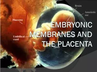
Histology of the Breast and Placenta
Histology of the breast Nipple - The breast is an organ containing 15-20 separate glands (mammary glands) each surrounded by connective tissue (stroma) - Functional gland (lactation) is a compound tubulo-acinar gland - Each mammary gland has its own duct (lactiferous duct) and sinus - Each duct empties independently at the nipple
Mammary glands Ducts Form majority of inactive gland Mostly, non-secretory Development regulated by estrogen Alveoli Derived from outgrowths of the ducts Present during pregnancy and lactation Cells synthesize and secrete milk Development regulated by progesterone
Functional stages of the breast Pre-puberty Puberty Adult Pregnancy Lactation Regression
Functional stages of the breast Pre-puberty Composed entirely of ducts, no alveoli Male and female structure similar Puberty Enlargement due to accumulation of adipose tissue Increase in duct system and degree of branching No alveoli present
Inactive (adult) Glands composed entire of ducts with minor alveolar development during mid to late menstrual cycle
Alveoli Alveoli Mid-Pregnancy Increased branching of ducts (estrogen) Alveoli develop from ducts (progesterone) Late Pregnancy Breast enlargement due to increased alveolar development Lumens of alveoli widen with accumulating milk
Alveoli Alveoli Lactation Alveolar cells actively synthesize and secrete milk Alveoli become widely distended with milk
Functional stages of the breast Inactive Pregnancy Lactating
Lactation Apocrine - lipid secretion Lipid droplet coalesce in cytoplasm, released as membrane bound vesicles containing some cytoplasm Merocrine - protein secretion Protein packaged in membrane-bound secretory vesicles, released by exocytosis Protein Lipid Lipid Lipid
Lactation Secretory products from alveolar cells Colostrum Secreted immediately after birth Protein rich High antibody titer (plasma cells in CT) Milk Increased lipid content over colostrum Contains protein, salts, carbohydrates, vitamins and antibodies Secretion starts several days after birth
Regulation of milk production and ejection High levels of estrogen and progesterone during pregnancy prevent milk production by blocking the action of prolactin Estrogen, Progesterone (before birth) X Neurohormonal suckling reflex arc Suckling initiates nerve impulses which regulate secretions from the pituitary Prolactin: Milk synthesis and secretion Oxytocin: Milk ejection
Functional stages of the breast Pre-puberty Puberty Adult Pregnancy Lactation Regression
Breast cancer Most common female cancer in the US; 1/3 of all cancer diagnoses Second most common cause of cancer death in women after lung cancer 12.5% (1 in 8) of all women born the United States will develop breast cancer at some time in their life 250,000 new cases each year, 40,000 deaths
Breast cancer Cancers arise from the ductal cells: Terminal ducts: Lobular carcinomas (20%) Larger ducts: Ductal carcinomas (80%) Types: Ductal carcinoma in situ (DCIS) (15-30%) Cannot penetrate the basal lamina and remains confined to the breast Generally not life threatening, though can become invasive if not treated Invasive ductal carcinoma (IDC) (70-85%) Readily metastasize outside the breast Contraindications for estrogen hormone replacement therapy
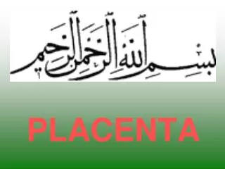
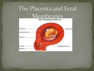
Placenta Transient organ formed by the fusion of fetal tissue, the chorion with the maternal uterine endometrium, the decidua At term 15-20 centimeters in diameter, 2-3 cm in thickness, 500-600 g 9-10 weeks 25-30% coverage
Placenta Functions: - Provides exchange of respiratory gases and nutrients and wastes between maternal and fetal circulations. Provides 90 sq. meters of surface area!!!! - Secretes hormones: Progesterone (progestins), estrogens, placental lactogens, and chorionic gonadotropins (hCG) - Transports macromolecules, e.g., IgG 9-10 weeks 25-30% coverage
(decidua) Blastocyst Stage of the embryo that implants into the uterus Composition: Trophoblast cells form the peripheral rim of the blastocyst cavity; invade the decidua and form the fetal portion of the placenta Inner cell mass, an eccentrically located cluster of cells inside the trophoblast which develops into the embryo
Chorion (fetal) Syncytio- trophoblast Cyto- tropho- blast Intervillous space Decidua (maternal) Trophoblast differentiates into: Cytotrophoblast (inner) Syncytiotrophoblast (outer) Vascular spaces form in the syncytiotrophoblast which connect with material circulation Cytotrophoblast grows out radially forming villi and laterally to enclose the intervillous spaces 16 days 21 days
Fetal side (Chorion) Stem villi Maternal side (Decidua) 100x 40x Inter- villous space
Syncytio- trophoblast Cyto- trophoblast Intervillous space Blood Chorionic villi Core of fetal connective tissue with fetal blood vessels Cytotrophoblast: Simple cuboidal epithelium; becomes discontinuous during late pregnancy Syncytiotrophoblast: Single cell (syncytium), multinucleated Covers the surface of the villus, facing the intervillous spaces and contacts maternal blood Possesses microvilli and abundant organelles associated with both protein and steroid hormone production Secretes a variety of hormones Chorionic plate Inter- villous space Decidua
The Decidua, the maternal placenta Decidua basalis – underlies the implanted conceptus forming the maternal portion of the placenta Decidua capsularis – covers the luminal surface of the conceptus; fuses with decidua parietalis, obliterating the uterine lumen Decidua parietalis - lines the remainder of the uterus
Umbilical cord V A A Placental Blood Flow Fetal Umbilical arteries (2) travel from the fetus through the umbilical cord to the placenta. Carry blood that is high in carbon dioxide and low in nutrient content Umbilical vein (1) returns oxygen-rich, nutrient-rich blood to the fetus Wharton’s jelly (mucus CT) Maternal Spiral arteries penetrate trophoblastic shell and spurt blood into the intervillous spaces, bathing the villi
Chorionic villus Intervillous space (Maternal blood) Placental interhemal barrier Separates maternal and fetal blood supplies, which normally do not mix. Beginning in the fetal capillary, this barrier consists of: - Capillary endothelial cell and its basement membrane - Fetal connective tissue of villus - Cytotrophoblast and its basement membrane - Syncytiotrophoblast
Placental interhemal barrier Separates maternal and fetal blood supplies, which normally do not mix. Beginning in the fetal capillary, this barrier consists of: - Capillary endothelial cell and its basement membrane - Fetal connective tissue of villus - Cytotrophoblast and its basement membrane - Syncytiotrophoblast
Placental interhemal barrier Separates maternal and fetal blood supplies, which normally do not mix. Beginning in the fetal capillary, this barrier consists of: - Capillary endothelial cell and its basement membrane - Fetal connective tissue of villus - Cytotrophoblast and its basement membrane - Syncytiotrophoblast
Placental interhemal barrier Separates maternal and fetal blood supplies, which normally do not mix. Beginning in the fetal capillary, this barrier consists of: - Capillary endothelial cell and its basement membrane - Fetal connective tissue of villus - Cytotrophoblast and its basement membrane - Syncytiotrophoblast
Placental interhemal barrier Separates maternal and fetal blood supplies, which normally do not mix. Beginning in the fetal capillary, this barrier consists of: - Capillary endothelial cell and its basement membrane - Fetal connective tissue of villus - Cytotrophoblast and its basement membrane - Syncytiotrophoblast