
Fertilization & Pregnancy
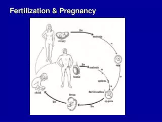
Fertilization & Pregnancy. of the 150-300 million sperm that are released upon ejaculation, only 100 will make it close to an egg.

Fertilization & Pregnancy
E N D
Presentation Transcript
of the 150-300 million sperm that are released upon ejaculation, only 100 will make it close to an egg. • the egg (ovum) is surrounded by 2 layers called the corona radiataand the zonapellucida, both of which must be penetrated by sperm before the egg is fertilized. • It takes between 3-5 for the egg to travel the 10-12 cm of the Fallopian Tube to the Uterus
embedded in the ovum membrane are receptors that participate in the binding of the sperm and the egg. Sperm may live 3-5 days in the female body.
once a sperm has attached, the receptors change shape to discourage other sperm from attaching to another receptor on the egg’s membrane (this prevents interspecific fertilization) • the acrosome of the sperm releases the enzyme (haluronidase), which digests a path through the corona radiata of the egg. • the “naked” sperm is able to penetrate the zona pellucida.
once the sperm has entered, the cortical reaction occurs. A surge in intracellular calcium activates the oocyte to prepare for cell division. It also causes the cortical granules, located just under the plasma membrane, to spill their contents into the extra cellular space beneath the zonapellucida.
the spilled material binds water, and as it swells, it detaches all sperm still in contact with the oocyte membrane, accomplishing the permanent slow block to polyspermy. In rare cases of polyspermy that do occur, the embryos contain too much genetic material and are nonviable (and die).
A fertilized egg is now called a ZYGOTE! Nucleii of egg and sperm fuse 23 chromosomes of ovum + 23 chromosomes of sperm = 46 chromosomes of zygote.
The zygote moves down the fallopian tube (oviduct), towards the uterus, dividing along the way. • After a few days, a morula is formed from the zygote. • A morula is a solid ball of cells (blastomeres) produced by cleavage. (Mitotic divisions; no cell growth so cells get smaller with each successive division)
fraternal twins would form if two eggs were released upon ovulation, and were both fertilized by sperm. • identical twins may occur at this point (because it is prior to cell differentiation), if the mitotic divisions cause the morula to split completely in two, or if one single cell separates from the morula and continues to divide.
Facts about Identical Twins The later in pregnancy that twinning occurs, the more structures will be shared. Zygotes that twin at the earliest stages will be diamniotic and dichorionic. Twinning between 4 to 8 days after fertilization typically results in monochorionic-diamniotic twins. Twinning between 8 to 12 days after fertilization will usually result in monochorionic-monoamniotic twins. Twinning after 12 days post-fertilization will typically result in conjoined twins.
Facts about Identical Twins Monozygotic twins are genetically identical unless there has been a mutation in development, and they are almost always the same gender. Examination of details such as fingerprints can tell them apart. As they mature, identical twins often become less alike because of lifestyle choices or external influences such as scars.
At day 5, 6, or 7 after fertilization (conception), the embryo is called a blastocyst, “a hollow ball of cells”; the blastocyst consists of a ring of outer cells (supporting cells) and a clump of cells at one end inside —the inner cell mass— which will become the embryo. Blastocyst
The disk of cells at one end will form the baby The cavity is the blastocoel The outer layer of cells will form the chorion which will form the placenta, the exchange site between baby and mom.
for pregnancy to continue, progesterone and estrogen levels must be maintained, and not drop, otherwise uterine contractions will occur and the embryo will be lost.
The choriongrows “fingers” called villi that grow into the endometrium about 5-6 days after fertilization. (Implantation) a. The chorion secretes HCG (human chorionic gonadotropin) which stimulates the corpus luteum to maintain the endometrium (More estrogen and progesterone). Keeps it around for approx. 3 months until the placenta is formed. b. HCG used in pregnancy test!
the blastocyst digests its way into the endometrium, and the embryo becomes surrounded by a pool of mother’s blood and tissue which supplies nutrients and removes wastes from the embryo by diffusion.
Stages of pregnancy There are nine months of pregnancy (~40 weeks from the date of the mother’s last menstrual period, or ~38 weeks from conception) and it is broken down into THREE trimesters.
1) The First Trimester • extends from fertilization to the end of the third month (12 weeks) - by the second week of development, three germ layers begin to form in the embryo (called gastrulation) . • the blastocyst becomes a gastrula, an embryo that has the 3 germ layers ectoderm, endoderm and mesoderm that are beginning to form.
During gastrulation, a split in the cell mass occurs to produce a space called the amniotic cavity or amnion. • Protects fetus from infection, dehydration, impact & changes in temp. The yolk sac. Produces blood cells, and nutrients for developing embryo in it’s early stages of life. Two layers of cells in the middle. This will become the baby.
The two layers of cells grow at different rates to form 3 cell layers.
The three embryonic cell layers are: a. Ectoderm - outer layer Forms skin, hair and nervous system. b. Mesoderm - middle layer Forms muscle, bone, blood, excretory and repro systems c. Endoderm - inner layer Digestive and respiratory organs.
The amnion will grow and the yolk sac will shrink. i. The amnion will surround the embryo to form the amniotic sac, a fluid filled sac. ii. Folding of the three cell layers makes a 3D baby.
at approximately week 5, diffusion becomes more and more inadequate for the needs of the embryo and the extra embryonic membranes begin to develop into the placenta. • the placenta is the site of exchange of nutrients and wastes between mother and fetus, and is fully developed by the end of the third month. • cells of the fetus and cells of the endometrium comprise the placenta.
The placenta sits against the uterine lining and is the exchange site between baby and mom.
the allantois membrane provides blood vessels for the placenta. • once the placenta has fully developed (2-3 months), the corpus luteum disintegrates and the placenta takes over in the release of estrogen and progesterone. • The umbilical cord connects the embryo to the placenta. Formed from body stalk, yolk sac & allantois. Contains 2 arteries which carry wastes away from baby. 1 vein that carries nutrients to baby.
2) Second Trimester - Weeks 13-27 1. All major organs formed. 2. Fetus becomes more human in shape. 3. Movement detected by mother 4. Cartilage becomes bone. 5. Grows from 50 to 350 mm+ 6. The placenta secretes progesterone to maintain pregnancy 7. A 27 week fetus may survive with special care.
3) Third trimester - 28- 40 weeks 1. Period of rapid growth --> mom gets huge! 2. Organ systems mature 3. Movements become strong and frequent 4. Baby turn head down due to gravity. Week 6 Week 16 Week 24 Week 32 Week 40
Sex Determination • The principle gene for sex determination is on the Y chromosome (from the father) • In the presence of the SRY gene testes will form • Read page 535