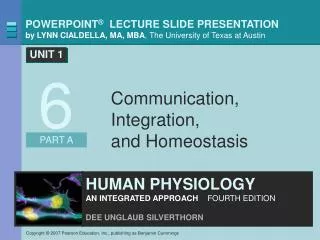
Communication, Integration, and Homeostasis
510 likes | 1.07k Vues
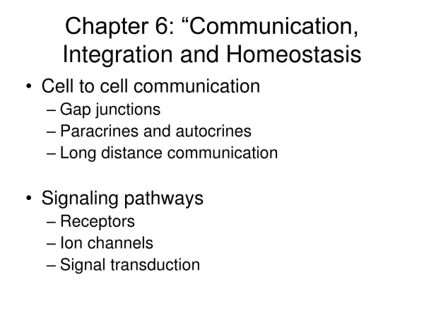
Communication, Integration, and Homeostasis. 6. About this Chapter. Cell-to-cell communication Signal pathways Novel signal molecules Modulation of signal pathways Control pathways Response loops Feedback loops. Cell-to-Cell Communication: Overview. Physiological signals

Communication, Integration, and Homeostasis
E N D
Presentation Transcript
About this Chapter • Cell-to-cell communication • Signal pathways • Novel signal molecules • Modulation of signal pathways • Control pathways • Response loops • Feedback loops
Cell-to-Cell Communication: Overview • Physiological signals • Electrical signals • Changes in cell’s membrane potential • Chemical signals • Secreted by cells into ECF • Responsible for most communication within the body • Target cells, or targets, receive signals • Four basic methods of communication
Cell-to-Cell Communication: Methods Direct contact and local cell-to-cell communication Gap junctions transfer both chemical and electrical signals Figure 6-1a
Cell-to-Cell Communication: Methods Direct contact and local cell-to-cell communication CAMs transfer signals in both directions Figure 6-1b
Cell-to-Cell Communication: Methods Paracrine and autocrine are chemical signals Figure 6-1c
Cell-to-Cell Communication: Methods Long distance cell-to-cell communication Figure 6-2a
Cell-to-Cell Communication: Methods Neurotransmitters have a rapid effect Figure 6-2b
Cell-to-Cell Communication: Methods Figure 6-2c
Signal Pathways: Overview Figure 6-3
Signal Pathways: Receptor locations Target cell receptors Figure 6-4 (1 of 2)
Signal Pathways: Receptor locations Figure 6-4 (2 of 2)
Signal Pathways: Membrane Receptors Four categories of membrane receptors Figure 6-5
Signal Pathways: Signal Amplification Transducers convert extracellular signals into intracellular messages which create a response Figure 6-7
Signal Pathway: Signal Transduction Steps of a cascade Steps of signal transduction pathway form a cascade Figure 6-9
Signal Pathway: Receptor Enzymes Tyrosine kinase, an example of receptor-enzyme Figure 6-10
Signal Pathway: GPCR • Membrane-spanning proteins • Cytoplasmic tail linked to G protein, a three-part transducer molecule • When G proteins are activated, they • Open ion channels in the membrane • Alter enzyme activity on the cytoplasmic side of the membrane
GPCR: Adenylyl Cyclase-cAMP Signal molecule binds to G protein-linked receptor, which activates the G protein. 1 One signal molecule G protein- coupled receptor 1 Adenylyl cyclase 2 G protein turns on adenylyl cyclase, an amplifier enzyme. 2 3 ATP G protein 3 Adenylyl cyclase converts ATP to cyclic AMP. cAMP 4 4 cAMP activates protein kinase A. Protein kinase A 5 5 Protein kinase A phosphorylates other proteins, leading ultimately to a cellular response. Phosphorylated protein Cell response The G protein-coupled adenylyl cyclase-cAMP system Figure 6-11
GPCR: Adenylyl Cyclase-cAMP Signal molecule binds to G protein-linked receptor, which activates the G protein. 1 One signal molecule G protein- coupled receptor 1 G protein Figure 6-11, step 1
GPCR: Adenylyl Cyclase-cAMP Signal molecule binds to G protein-linked receptor, which activates the G protein. 1 One signal molecule G protein- coupled receptor 1 Adenylyl cyclase 2 G protein turns on adenylyl cyclase, an amplifier enzyme. 2 G protein Figure 6-11, steps 1–2
GPCR: Adenylyl Cyclase-cAMP Signal molecule binds to G protein-linked receptor, which activates the G protein. 1 One signal molecule G protein- coupled receptor 1 Adenylyl cyclase 2 G protein turns on adenylyl cyclase, an amplifier enzyme. 2 3 ATP G protein 3 Adenylyl cyclase converts ATP to cyclic AMP. cAMP Figure 6-11, steps 1–3
GPCR: Adenylyl Cyclase-cAMP Signal molecule binds to G protein-linked receptor, which activates the G protein. 1 One signal molecule G protein- coupled receptor 1 Adenylyl cyclase 2 G protein turns on adenylyl cyclase, an amplifier enzyme. 2 3 ATP G protein 3 Adenylyl cyclase converts ATP to cyclic AMP. cAMP 4 4 cAMP activates protein kinase A. Protein kinase A Figure 6-11, steps 1–4
GPCR: Adenylyl Cyclase-cAMP Signal molecule binds to G protein-linked receptor, which activates the G protein. 1 One signal molecule G protein- coupled receptor 1 Adenylyl cyclase 2 G protein turns on adenylyl cyclase, an amplifier enzyme. 2 3 ATP G protein 3 Adenylyl cyclase converts ATP to cyclic AMP. cAMP 4 4 cAMP activates protein kinase A. Protein kinase A 5 5 Protein kinase A phosphorylates other proteins, leading ultimately to a cellular response. Phosphorylated protein Cell response Figure 6-11, steps 1–5
GPCR: The Phospholipase C System Signal molecule Extracellular fluid 1 Membrane phospholipid Cell membrane 3 2 4 PL-C DAG PK-C Intracellular fluid Protein + Pi Receptor IP3 G protein 5 KEY Phosphorylated protein PL-C = phospholipase C DAG = diacylglycerol PK-C = protein kinase C IP3 = inositol trisphosphate ER = endoplasmic reticulum Ca2+ stores Ca2+ ER Cellular response 1 2 3 4 5 DAG activates protein kinase C (PK-C), which phosphorylates proteins. IP3 causes release of Ca2+ from organelles, creating a Ca2+ signal. Signal molecule activates receptor and associated G protein. G protein activates phospholipase C (PL-C), an amplifier enzyme. PL-C converts membrane phospholipids into diacylglycerol (DAG), which remains in the membrane, and IP3, which diffuses into the cytoplasm. Figure 6-12
GPCR: The Phospholipase C System Signal molecule Extracellular fluid 1 Cell membrane Intracellular fluid Receptor G protein KEY PL-C = phospholipase C DAG = diacylglycerol PK-C = protein kinase C IP3 = inositol trisphosphate ER = endoplasmic reticulum 1 Signal molecule activates receptor and associated G protein. Figure 6-12, step 1
GPCR: The Phospholipase C System Signal molecule Extracellular fluid 1 Cell membrane 2 PL-C Intracellular fluid Receptor G protein KEY PL-C = phospholipase C DAG = diacylglycerol PK-C = protein kinase C IP3 = inositol trisphosphate ER = endoplasmic reticulum 1 2 Signal molecule activates receptor and associated G protein. G protein activates phospholipase C (PL-C), an amplifier enzyme. Figure 6-12, steps 1–2
GPCR: The Phospholipase C System Signal molecule Extracellular fluid 1 Membrane phospholipid Cell membrane 3 2 PL-C DAG Intracellular fluid Receptor IP3 G protein KEY PL-C = phospholipase C DAG = diacylglycerol PK-C = protein kinase C IP3 = inositol trisphosphate ER = endoplasmic reticulum 1 2 3 Signal molecule activates receptor and associated G protein. G protein activates phospholipase C (PL-C), an amplifier enzyme. PL-C converts membrane phospholipids into diacylglycerol (DAG), which remains in the membrane, and IP3, which diffuses into the cytoplasm. Figure 6-12, steps 1–3
GPCR: The Phospholipase C System Signal molecule Extracellular fluid 1 Membrane phospholipid Cell membrane 3 2 4 PL-C DAG PK-C Intracellular fluid Protein + Pi Receptor IP3 G protein KEY Phosphorylated protein PL-C = phospholipase C DAG = diacylglycerol PK-C = protein kinase C IP3 = inositol trisphosphate ER = endoplasmic reticulum Cellular response 1 2 3 4 DAG activates protein kinase C (PK-C), which phosphorylates proteins. Signal molecule activates receptor and associated G protein. G protein activates phospholipase C (PL-C), an amplifier enzyme. PL-C converts membrane phospholipids into diacylglycerol (DAG), which remains in the membrane, and IP3, which diffuses into the cytoplasm. Figure 6-12, steps 1–4
GPCR: The Phospholipase C System Signal molecule Extracellular fluid 1 Membrane phospholipid Cell membrane 3 2 4 PL-C DAG PK-C Intracellular fluid Protein + Pi Receptor IP3 G protein 5 KEY Phosphorylated protein PL-C = phospholipase C DAG = diacylglycerol PK-C = protein kinase C IP3 = inositol trisphosphate ER = endoplasmic reticulum Ca2+ stores Ca2+ ER Cellular response 1 2 3 4 5 DAG activates protein kinase C (PK-C), which phosphorylates proteins. IP3 causes release of Ca2+ from organelles, creating a Ca2+ signal. Signal molecule activates receptor and associated G protein. G protein activates phospholipase C (PL-C), an amplifier enzyme. PL-C converts membrane phospholipids into diacylglycerol (DAG), which remains in the membrane, and IP3, which diffuses into the cytoplasm. Figure 6-12, steps 1–5
Signal Pathway: Receptor-Channel Extracellular signal molecules Ions Receptor-channels open or close in response to signal molecule binding. 1 1 G protein- coupled receptor Ion channel 2 Some channels are directly linked to G proteins. 2 G protein Other ligand-gated channels respond to intracellular second messenger. 3 Change in membrane permeability to Na+, K+, Cl– 3 Intracellular signal molecules Creates electrical signal Voltage-sensitive protein Cellular response How ions create electrical signals Figure 6-13
Signal Pathway: Receptor-Channel Extracellular signal molecules Ions Receptor-channels open or close in response to signal molecule binding. 1 1 Ion channel Figure 6-13, step 1
Signal Pathway: Receptor-Channel Extracellular signal molecules Ions Receptor-channels open or close in response to signal molecule binding. 1 1 G protein- coupled receptor Ion channel 2 Some channels are directly linked to G proteins. 2 G protein Figure 6-13, steps 1–2
Signal Pathway: Receptor-Channel Extracellular signal molecules Ions Receptor-channels open or close in response to signal molecule binding. 1 1 G protein- coupled receptor Ion channel 2 Some channels are directly linked to G proteins. 2 G protein Other ligand-gated channels respond to intracellular second messenger. 3 3 Intracellular signal molecules Figure 6-13, steps 1–3
Signal Pathway: Receptor-Channel Extracellular signal molecules Ions Receptor-channels open or close in response to signal molecule binding. 1 1 G protein- coupled receptor Ion channel 2 Some channels are directly linked to G proteins. 2 G protein Other ligand-gated channels respond to intracellular second messenger. 3 Change in membrane permeability to Na+, K+, Cl– 3 Intracellular signal molecules Creates electrical signal Voltage-sensitive protein Cellular response Figure 6-13
Signal Pathway: Signal Transduction Summary map of signal transduction systems Figure 6-14