Download
1 / 23
260 likes | 838 Vues
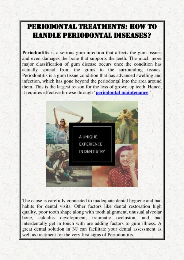
Ch. 24 Periodontal diseases. Introduction. Periodontal disease includes all disorders of the supporting structures of the teeth, namely the gingiva , periodontal ligament and supporting alveolar bone .

E N D
Introduction • Periodontal disease includes all disorders of the supporting structures of the teeth, namely the gingiva, periodontal ligament and supporting alveolar bone. • This may cause gingivitis or severe inflammation of the periodontal ligament called periodontitis • They are associated with a shift in the balance of the resident microflora, a similar situation to dental caries
Gingivitis • Plaque-associated gingivitis - The microflora of the healthy gingiva composed mainly of Gram-positive cocci, especially Streptococcus spp. - Toxins released by these bacteria induce an inflammatory response in the gingival tissues - Clinically, gingivitis is characterized by redness, gingival bleeding and edema. • Acute ulcerative gingivitis is a specific form of gingivitis in which there is necrosis of the tips of the gingival papillae, spontaneous bleeding • Atypical gingivitis occurs in immunocompromised as acute leukemia and HIV.
Stages in the development of gingivitis • The initial lesion: -This develops within 4 days of plaque accumulation. consists mostly of Gram-positive cocci (Streptococcus spp). there is an acute inflammatory reaction. Clinically, it is not visible • The early lesion: - Appears after 7 days of plaque accumulation and is detectable clinically as gingivitis. Actinomyces spp., spirochaetes and capnophilic organisms are now more due to the lower O2 tension. The most inflammatory infiltrated cells are lymphocytes (75%) and macro-phages, with some plasma cells.
Stages in the development of gingivitis • Established lesion: • Growth of obligate anaerobes such as Porphyromonas gingivalis and Prevotella intermedia started. • A periodontal pocket lined with pocket epithelium may be present • Established lesions may persist for months or years without progression to periodontitis
Periodontitis • Defined clinically as inflammation of the supporting tissues of the teeth • It is as established lesion of gingivitis, with additional migration of the junctional epithelium down the root surface, alveolar bone resorption and subsequent pocket formation • It is also characterized by progressively destructive changes which destroy alveolar bone and periodontal ligament • Formation of a periodontal pocket creates an environment that is highly anaerobic
The etiology of periodontal disease • Host and microbial factors 1. Host factors : Some of the factors which may increase host susceptibility to infection include inadequate or unregulated host immune response, diabetes mellitus, stress and tobacco use. Host factors include Igs, Complement, Cytokines, PMNs & Macrophages, T & B cells.
The etiology of periodontal disease 2. Microbial factors: Attachment to host tissue, multiplication at the suspected site, evasion of host defences, enzymes,endotoxin, proteases of IgG & IgA, leukotoxins, superoxide dismutase
Diagnosis • Bacteria present in periodontal pockets may be detected by microbial culture techniques, detection of certain microbial enzymes, immunological methods and DNA/RNA probes
Treatment of periodontal disease • supragingival plaque control • root surface debridement • surgery, if improved access is required • consideration of adjunctive antimicrobial agents
Ch. 25Infections of the pulp, periapical tissues and bone of the jaw
Bacteria are responsible for both dental caries and periodontal diseases • Extension of these diseases commonly causes infection in the adjacent tissues, notably the pulp, periapical area and oro-facial soft tissues • It may infect the bone of the jaw to cause osteomyelitis
Pulpitis • Inflammation of the pulp may follow exposure to thermal, mechanical or chemical stimuli, in addition to microorganisms. • The most common cause of pulpal necrosis is dental caries, other factors include accidental trauma to the tooth, exposure of the pulp during instrumentation and spread of infection from a deep periodontal pocket.
Pulpitis • Pulpitis following dental caries • Pulpitis through an open cavity • Pulpitis through the apical foramen • Pulpal necrosis
Dentoalveolar abscess • Develops typically at the apices of the roots of teeth, following necrosis of the pulp • Dentoalveolar abscesses are endogenous infections usually caused by a mixture of bacteria including obligate anaerobes. • Pus specimens from the head and neck region for microbiological examination should be collected by needle aspiration • Drainage of pus is the essential element of treatment for a dentoalveolar or periodontal abscess.
Ludwig's angina • Ludwig's angina is a bilateral infection of the sublingual and sub-mandibular spaces. • The infection often represents cellulitis of the fascial spaces, rather than true abscess formation • Ludwig's angina is a life-threatening infection involving the sublingual and submandibular spaces • Maintenance of the airway is paramount in the management of Ludwig's angina
Osteomyelitis of the jaws • Osteomyelitis of the jaws is uncommon, but typically occurs in patients with deficient host defenses or reduced vascularity of the bone • Staphylococci were implicated as important organ-isms in osteomyelitis of the jaws, but also anaerobic Gram-negative rods and anaerobic streptococci are important • Osteomyelitis of the jaws is usually a mixed infection, requiring both medical and surgical treatment.
Actinomycosis • Actinomycosis is an endogenous infection, associated with Actinomyces israelii, which presents as a swelling, often at the angle of the mandible • The disease is usually a chronic, long-standing infection • It presents typically as a swelling, often at the angle of the lower jaw • “Sulphur granules” are particles seen in pus from actinomy-cotic lesions and which contain aggregates of actinomyces filaments • Actinomycosis is treated by surgical drainage and long-term administration of antibiotics, ideally penicillin
Salivary gland infections • Salivary gland infections may be viral or bacterial • It may be acute or chronic • The most common cause is mumps virus, but acute sialadenitis is usually bacterial in origin. • Many factors are involved in the pathogenesis of salivary gland infections as dehydration, previous radiotherapy, sialolithiasis
Viral infections of salivary glands • Mumps: - Mumps virus is transmitted by direct contact with saliva and droplet spread. - Mumps is characterized by inflammation and enlargement of the salivary glands. - A clinical diagnosis of mumps can be confirmed by detection of specific IgM • Human immunodeficiency virus - HIV infection may cause xerostomia and enlargement of the major salivary glands
Bacterial infections of salivary glands • Bacterial parotitis normally occurs in patients with reduced salivary flow or abnormalities in gland architecture • The swelling of bacterial parotitis is exquisitely painful and pus may be expressed from the duct opening • It may be acute or chronic • Treatment of acute bacterial parotitis is by hydration, antibiotics and, if necessary, surgical drainage
Bacterial infections of salivary glands • Following resolution of acute parotitis, sialography should be undertaken to identify correctable salivary gland abnormalities. • Recurrent parotitis of childhood should be treated conservatively, since most cases resolve spontaneously at puberty. • Tuberculosis and Actinomycosis are less common bacterial causes of salivary-gland infection.