Advanced DTI and Shape Analysis Tools for Longitudinal Brain Imaging Studies
Explore cutting-edge tools and methodologies developed at UNC and Utah for analyzing Diffusion Tensor Imaging (DTI) and shape differences in brain structures. This comprehensive framework allows for high-sensitivity localized analysis and addresses various artifacts and errors in DTI data. Key achievements include over 50 clinical and methodological papers and innovative Slicer extensions for enhanced user support. The training provided fosters community development, leading to further applications in brain pathology, segmentation, and statistical analysis, crucial for understanding brain development and disorders.

Advanced DTI and Shape Analysis Tools for Longitudinal Brain Imaging Studies
E N D
Presentation Transcript
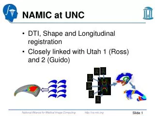
NAMIC at UNC DTI, Shape and Longitudinal registration Closely linked with Utah 1 (Ross) and 2 (Guido)
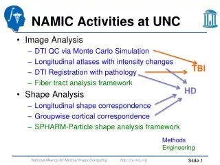
UNC-Utah DTI Fiber Analysis Analysis of DTI properties along the fiber Many years of methods & tool development Allows for localized analysis with high sensitivity
UNC DTI Activities DTI QC: Vibration artifacts, Error estimation Fibers: Post-processing, clustering Statistical analysis methods Main tool development, Slicer extensions, batch and grid processing
DTI Achievements AJP, 12 Cerebral Cortex, 12 • First “true” fiber based analysis • >25 clinical papers • Designed for human data, used for primate and rodent MRI’s • >25 methodological papers • Slicer extensions for investigators • SPIE tutorials • Training @ UNC • Framework only available within last year => expect many more papers from the community
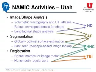
Cortical Correspondence • Goal: Flexible, group-wise cortex correspondence • Cortical thickness analysis in HD DBP • Allow for point and sulcal landmarks, longitudinal info • Existing NA-MIC particle based correspondence • No guarantee on surface mesh topology • Spherical parametrization • No explicit registration/deformation • Spherical harmonics encoding • Local angular deformation • Optimal pole choice
UNC-Utah Shape Tools • Analysis of shape differencesor changes • Pathology in brain structures, Segmentation QC studies, bone structures in Paleontology, Botany & Anthropology • Localization of pathology, classification via shape • Comprehensive toolset: classical SPHARM-PDM, implied medial description, group-wise particle description Volumetric analysis: Size, Growth Statistical analysis Shape Representation Binary Segmentation
UNC Shape Activities • Particle based correspondence for complex and non-spherical topology cases • Convoluted (cortex) and narrow (mandible)surfaces • Incorporating longitudinal modeling • Implied medial description • Statistical analysis methods • Main tool development • Slicer extensions, batch and grid processing, visualization tools
Shape Achievements Neuron, 13 OSOMOPORE, 11 NeuroImage, 12 NeuroImage, 13 • >25 clinical papers with NA-MIC co-authors • >20 clinical papers without NA-MIC co-authors • >25 methodological papers • Slicer extensions for investigators • Training @ UNC • No other (non-technical) shape analysis toolset around • SPHARM considered standard, new methods compare to it
Longitudinal Atlas Building Splenium 3D atlas 4D atlas 4D atlas with individual growth/change trajectories For DTI and structural MRI
4D Intensity Changes Average A-P intensity change in WM • Models intensity changes across time • logistic function, model is incorporated into intensity match • Handles non-balanced & missing samples • Allows for joint processing of longitudinal intra-subject data • Structural Segmentation & tissue classification • Enhances stability => sensitivity to detect longitudinal changes
Intensity Change Images Individual (red), median (blue) logistic curves, image intensity (dots) for 2 voxels Thinned white matter mask • Intensity change “atlas image” as byproduct • Estimated onset (in months) and rate of maturation
NA-MIC @ UNC DTI & Shape & Longitudinal Registration Field leading tools (Slicer extensions) for investigators, not just engineers Training support Leading to high number of papers & applications