Download
1 / 10
100 likes | 309 Vues
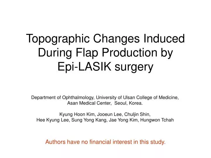
Topographic Changes Induced During Flap Production by Epi-LASIK surgery. Department of Ophthalmology, University of Ulsan College of Medicine, Asan Medical Center, Seoul, Korea. Kyung Hoon Kim, Jooeun Lee, Chuljin Shin, Hee Kyung Lee, Sung Yong Kang, Jae Yong Kim, Hungwon Tchah.

E N D
Topographic Changes Induced During Flap Production by Epi-LASIK surgery Department of Ophthalmology, University of Ulsan College of Medicine, Asan Medical Center, Seoul, Korea. Kyung Hoon Kim, Jooeun Lee, Chuljin Shin, Hee Kyung Lee, Sung Yong Kang, Jae Yong Kim, Hungwon Tchah Authors have no financial interest in this study.
Introduction • Changes in corneal topography and ocular aberration are highly evaluated as a measurement of visual quality after refractive surgery. • Currently, customized surface ablation techniques are more frequently used to improve patients quality of vision. • However, these refractive procedures are conducted under the assumption that removed corneal epithelium does not significantly affect outcomes of refractive surface ablation and that they will be regenerated into their original distribution. • Thus, the purpose of this study was to investigate induced changes of corneal topographic data before and after epithelial flap removal during Epi-LASIK.
Patients and Methods • Patients • Eighteen eyes of ten subjects undergoing Epi-LASIK for correction of myopia or myopic astigmatism were enrolled. • Surgical Technique • Microkeratome (Amadeus II®, AMO, Irvine, CA) for epithelial flap production. • Flap diameter : 9.0mm • Flap thickness : Approximately 55~60µm • Topography • Corneal topography with ray tracing aberrometry, i-Trace® (Technologies, Houston, Texas), was used to measure corneal topography before and after epithelium removal. • Corneal aberrations are calculated using the program integrated in the i-Trace system based on the topographic data. Fig.1 i-Trace® Fig.2 Amadeus microkeratome®
Results (1) • Simulated K showed statistically meaningful change between the epithelium on and off state. • Tendency of Increasing refractive power after epithelium removal. • However, other topographic parameters (central power, astigmatism on simulated K, asphericity and best fit sphere) did not significantly change. Fig.3 Topography with epithelium Fig.4 Topography without epithelium
Total Corneal Higher Order Aberrations 3mm 5mm 7mm With Epithelium Without Epithelium Results (2) • Total Corneal higher order aberrations (HOAs) significantly increased at 3.0, 5.0, and 7.0mm diameters after epithelial removal.
Discussion (1) • According to Damien et al, epithelial distribution on the corneal surface before and after its removal during PRK is closely related to astigmatism and changes in corneal irregularity. They reported a trend for increased anterior prolateness after epithelial removal, which agrees with results of previous reports. • In this study, however, neither astigmatism nor anterior prolateness were significantly altered. Differing methods of epithelial removal may underlie such a result. • Increases of simulated K may be due to thicker epithelium in the periphery than in the center.
Discussion (2) • Increases of total HOAs after the removal of epithelium may indicate its role to smoothe the corneal surface over the Bowman’s layer and to decrease the corneal total HOAs. • However, current customized surface ablation is conducted in the epithelium off state whereas pre-operative wavefront data are measured from epithelium on normal cornea. This assumption that the epithelium does not affect significantly on refractive surface ablation may be erroneous. • For this reason, customized techniques in surface ablation may cause unpredictable results. Therefore, more concern should be given to changes of HOAs attributable to the corneal epithelium, and further study is needed to analyze causes in increase of HOAs.
Conclusions • Astigmatism of simulated K in corneal topography showed a tendency to increase after removing the epithelial flap during Epi-LASIK. • Total corneal HOAs at 3.0, 5.0, and 7.0mm increased after epithelial removal. • This suggests that the corneal epithelium could have a significant effect on corneal topography and the results of corneal surface ablation. • Future surface ablation refractive surgery may require more concern and investigation into the role of the corneal epithelium.
References 1. Damien G, Louis R, Thanh H-X. Contribution of the corneal epithelium to anterior corneal topography in patients having myopic photorefractive keratectomy. J Cataract Refract Surg 2007; 33:1860-1865 2. Serrao S, Lombardo M. Corneal epithelial healing after photorefractive keratectomy: analytical study. J Cataract Refract. Surg 2005; 31: 930-937 3. Simon G, Ren Q, Kervick GN, Parel J-M. Optics of the corneal epithelium. Refract Corneal Surg 1993; 9:42-50 4. Patel S, Marshall J, Fitzke FW III. Refractive index of the human corneal epithelium and stroma. J Refract Surg 1995; 11:100-105 5. Corneal Topography in the Wavefront Era Wang M, Swartz T. Corneal Topography in the Wavefront Era; a Guide for Clinical Application. Thorofare, SLACK Incorporated, 2006