Download
1 / 25
250 likes | 421 Vues
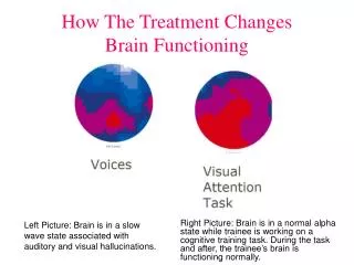
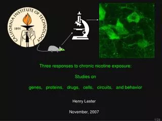
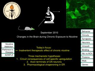
Behavior. September 2010. Changes in the Brain during Chronic Exposure to Nicotine. Circuits. Synapses. Neurons. Nicotine Addiction. Intracell. Binding. Parkinson’s Disease. Today’s focus: Inadvertent therapeutic effect of chronic nicotine Three mechanistic hypotheses

E N D
Behavior September 2010 Changes in the Brain during Chronic Exposure to Nicotine Circuits Synapses Neurons Nicotine Addiction Intracell. Binding Parkinson’s Disease • Today’s focus: • Inadvertent therapeutic effect of chronic nicotine • Three mechanistic hypotheses • Circuit consequences of cell-specific upregulation • Axon terminals of DA neurons • Pharmacological chaperoning in ER Nic vs ACh ADNFLE Proteins RNA Genes
1st and 2nd hypotheses. Cellular and axon/soma specificity of SePhaChARNS: α4* nAChRs CA EC MH DG IPN Medial Perforant Path Striatum SNc, VTA Thalamus, superior colliculus SNr, VTA Nashmi et al J Neurosci 2007; Xiao et al, J. Neurosci 2009
1st Hypothesis for PD neuroprotection by Chronic nicotine: Circuit-based mechanism in substantia nigra via Cholinergic, Dopaminergic, and GABAergic neurons in Hindbrain & Midbrain . . . Analogous to “deep brain stimulation” in subthalamic nucleus? STN Striatum SNc DAergic PPTg GABAergic neurons have increased (or more regular?) firing in chronic nicotine. . . Thalamus, superior colliculus Cholinergic GABAergic SNr Endogenous ACh Upregulated a4* nAChRs
β2 subunits govern the ER localization of α4β2 nAChRs α4-eGFP β2 5 µm α4-eGFP β4 4
Nicotine and mutant β2 subunits overcome a rate limiting ER exit step in α4β2 nAChR trafficking to the PM LFM AAQA 10 µm • Mutations overcome a rate limiting step in ER export • Nicotine upregulates nAChRs by a distinct mechanism 5
“Endoplasmic reticulum stress” occurs when the cell cannot clear newly synthesized proteins from the ER GTPase: Sar1 COP II: Sec23/24 heterodimer scission ER lumen ER membrane Cytosolic compartment Golgi complex Endoplasmic reticulum Plasma membrane Early endosome COPI ERGIC Clathrin COPII COPI nAChR nAChR Secretory vesicle Mancias & Goldberg, Traffic 2005
3rd hypothesis: Nicotine and mutant nAChRs increase ER exit sites 10 µm • M3-M4 mutations increase ER exit of nAChRs • Nicotine exposure increases ER exit of wt nAChRs • Upregulation is initiated prior to ER exit of nAChRs 7
Nicotine and mutant receptors alter nAChR stoichiometry in the ER 1.06 2.7 0.57 4.83 2.34 3.8 2.26 4.6 2.05 2.3 1.7 2.87 8
Nicotine and DM receptors alter trans-Golgi network activity GalT-mcherry, No Nic α4-eGFP β2-wt + 0.1 µM Nic α4-eGFP β2-wt, No Nic GalT-mcherry + 0.1 µM Nic GalT-mcherry α4-eGFP β2-DM Merge 9
Specific Expression of a6 nAChRs in Midbrain DA Neurons DA Neurons GABA Neurons WT a6 L9′S a4 L9′A 10
a4 Subunits are Required for Behavioral Hyperactivity Observed in a6 L9’S Mice 11 Ryan Drenan, Sheri McKinney Drenan et al., in preparation 2010
Conclusion: a4 Subunits are Critical to a6 nAChR Function In Vivo 12
2nd hypotheses (DA terminals). α4* nAChRs on dopaminergic terminals exert a tonic inhibition of glutamate release. In chronic nicotine, this tonic inhibition is greater because of upregulated α4* nAChRs This could be neuroprotective (Xiao et al, 2009).
Reliable α6β2 expression in N2a cells B A Vm = -60 mV, 0.3 mM ACh (0.1 s puff) Vm = -60 mV Vm = -90 mV 30 60 1.93 pA → 21.4 pS 1.44 pA → 24 pS 20 40 3 pA Counts 15 ms 10 20 0 0 -2.0 -1.5 -1.0 -0.5 -3.0 -2.5 -2.0 -1.5 -1 C Amplitude (pA) Amplitude (pA) D
Chronic nicotine exposure downregulates PM α6-meGFP β2 nAChRs ER subtracted No Nic + 0.1 µM Nic ER subtracted
α5 an “accessory” subunit • Forms receptors with α3β4 and α4β2 subunits • Not thought to contribute to ligand binding interface • Smallest intracellular loop (~70aa) • Regulated at the level of brain region, cell type and possibly mRNA • GWAS and candidate gene studies identified a SNP in the gene Chrna5. This Aspartic Acid to Asparagine mutation at position 398 appears to confer an increased risk for nicotine addiction (D398N) • KO mice show differences in nicotine self administration compared to wt mice • α5* receptors are not thought to upregulate when exposed to nicotine
Two stoichiometries of α4β2 receptors β2 β2 α4 α4 β2 α4 α4 β2 α4 β2 One stoichiometry of α4β2α5 receptors? α4 β2 β2 α5 α4
HEK 293 1 3 4 2 500ng ea. 6 8 5 7 • 1. α4GFPβ2 • 2.α4GFPβ2 α5stitzel • 3.α4β2 α5stitzel GFP385 • 4.α4β2 α5stitzel GFP385L • 5.α4β2 α5stitzel GFP378L • 6.α4β2 α5-IDT GFP385L • 7.α4β2 α5-IDT GFP378L • 8.α4β2 α5-IDT C-Term GFP • α4β2 α5-IDT D/N C-Term GFP • α4β2 β3P379 YFP 9 10
Moving Forward with α5. . . • Transfection of α5-GFP into HEK293 cells • NFRET studies yield information about receptor assembly • TIRF studies yield information about surface trafficking • Transfection of α5-GFP into mouse neurons • Live and fixed cell confocal imaging of α5-GFP localization in neurons All experiments will compare α5-GFP and α5D398N-GFP under + and - nicotine conditions for changes in receptor behavior
Lynx proteins modulate (inhibit) nicotinic receptor function 1-2Lynx proteins are GPI-linked variants of -neurotoxins (e.g. -bungarotoxin) 1Removal of lynx using genetically engineered mice cause nicotinic receptor hypsersensitivity and enhanced learning and memory 3 • Miwa et al., Neuron, 1999 • Ibanez-Tallon et al., Neuron, 2002 • 3. Miwa et al., Neuron, 2006 lynx btx
-bungarotoxin binds to nAChRs at subunit interfaces, suggesting a possible site of action of lynx on the receptor. • lynx proteins are GPI-linked membrane proteins. • GPI-linked protein sort to specialized lipid domains (e.g. rafts). • Lipid rafts have been shown to stabilize nicotinic receptors within synaptic structures, and reduce receptor mobility. • Therefore, lynx:nAChR interactions may cause receptors to localize to specialized domains and reduce receptor mobility. btx and AChBP
Questions:Do nAChR co-localize with lipid rafts? Labeling rafts by incubating transfected cells with labeled cholera toxin 1) co-labeling: 4b2 receptors fused in-frame to gfp/cherry 2) co-labeling: 7 receptors with labeled -bungarotoxin Does disruption of lipid rafts increase receptor mobility?Image cells with transfected receptor (in-frame labeled). Deplete cholesterol in rafts with methyl-b –cyclodextran) Does removal of lynx (e.g. lynx1KO mice) alter the mobility, trafficking pathways, or final localization, of nAChRs? Image primary neuronal cultures from wt vs. ko mice, transfected with labeled receptor
Differential effects of nicotinic ligands on the assembly, ER exit and PM localization of nAChRs • NFRET using α4-mcherry + β2-eGFP • Nicotine assembles (α4)2(β2)3 nAChRs • Cytisine assembles (α4)3(β2)2 nAChRs • Both drugs affect assembly in the ER 23
Differential effects of nicotinic ligands on the assembly, ER exit and PM localization of nAChRs Merge No Drug Sec24D-eGFP + 0.1 µM Nic, 48 h 4-mCherry + 2-eGFP + 0.1 µM Cyt, 48 h 10 µm 24
Differential effects of nicotinic ligands on the assembly, ER exit and PM localization of nAChRs Merge No Drug Sec24D-eGFP + 0.1 µM Nic, 48 h 4-mCherry + 2-eGFP 10 µm 25