Download
1 / 39
500 likes | 1.04k Vues
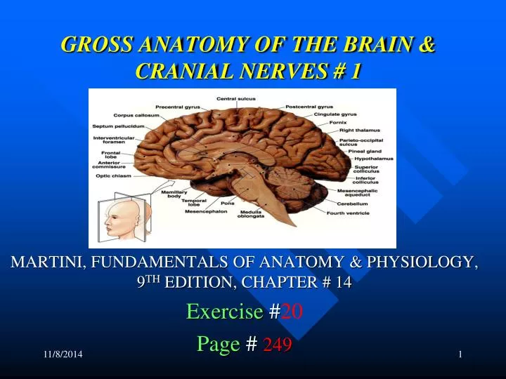
GROSS ANATOMY OF THE BRAIN & CRANIAL NERVES # 1. MARTINI, FUNDAMENTALS OF ANATOMY & PHYSIOLOGY, 9 TH EDITION, CHAPTER # 14 Exercise # 20 Page # 249. NOTE:. THIS IS A STUDY GUIDE , NOT AN ALL INCLUSIVE REVIEW.

E N D
GROSS ANATOMY OF THE BRAIN & CRANIAL NERVES # 1 MARTINI, FUNDAMENTALS OF ANATOMY & PHYSIOLOGY, 9TH EDITION, CHAPTER # 14 Exercise #20 Page#249
NOTE: • THIS IS A STUDY GUIDE, NOT AN ALL INCLUSIVE REVIEW. • THERE MIGHT BE THINGS NOT COVERED BY THIS STUDY GUIDE THAT MIGHT BEASKED IN YOUR PRACTICUMS / QUIZZES. • STUDENTS ARE RESPONSIBLE FOR READING THEIR TEXBOOK (S) AND FOR ALL THE MATERIAL COVERED DURING THE LABORATORY PERIOD, AS PER THE COURSE SYLLABUS
OBJECTIVES • Identifying the major regions of the brain and state their function. • Identifying the meninges. • Locating and naming selected cranial nerves.
CENTRAL NERVOUS SYSTEM (CNS)- BRAIN & SPINAL CORD • PERIPFERAL NERVOUS SYSTEM (PNS)- NERVES & ASSOCITED • GANGLIA • CEREBRAL HEMIPHERES- 2 HALF OF THE CEREBEUM, RIGHT & LEFT • DIVIDED BY LONGITUDINAL FISSURE • GYRI(PLURAL)- THICK AREAS • GYRUS- (SINGULAR)- FOLDS • PRECENTRAL GYRUS • POSTCENTRAL GYRUS • FISSURES: DEEP GROOVES • SULCI- SHALLOW GROOVES SEPARATING GYRI • SULCY- PLURAL • SULCUS- SINGULAR • LONGITUDINAL FISSURE- IT SEPARATES THE RIGHT & LEFT • HEMISPHERES • CENTRAL SULCUS- IT DIVIDES THE CEREBRAL HEMISPHERS IN • ANTERIOR & POSTERIOR
LATERAL SULCUS- IT SEPARATES FRONTAL AND PARIETAL LOBES • FROM TEMPORAL LOBE • PARIETO OCCIPITAL SULCUS- IT SEPARATES THE PARIETAL LOBE • FROM OCCIPITAL LOBE • FRONTAL LOBE- FROM CENTRAL SULCUS ANTERIORLY • PREFRONTAL CORTEX- MOST ANTERIOR PART OF THE FRONTAL LOBE • F- CENTER OF THE INTELECT FOR RAZONALIZATION • TEMPORAL LOBE- UNDER THE TEMPORAL BONES UNDER THE • LATERAL SULCUS • PARIETAL LOBE- UNDER THE PARIETAL BONES & BEHIND THE • FRONTAL LOBE • OCCIPITAL LOBE- POSTERIOR LOBE OF THE CEREBRUM • UNDER OCCIPITAL BONE
PRE CENTRAL GYRUS- GYRUS BEFORE THE CENTRAL SULCUS • (ORANGE IN MODEL) • IT CONTEINS THE PRIMARY MOTOR AREA- • F- TO CONTROL VOLUNTARY MUSCLE MOVEMENT • POSTCENTRAL GYRUS- GYRUS AFTER THE CENTRAL SULCUS • (LAVANDER IN MODEL) • IT CONTEIS THE PRIMARY SOMATOSENSORY AREA • F- TO RECEIVE SENSORY INFO FROM GENERAL • SENSORY RECEPTORS EX- TOUCH, PAIN, TEMP • SOMATOSENSORY ASSOCIATION AREA- • BEHIND THE POST CENTRAL GYRUS (LAVANDER) • F- TO INTERPRET SENSORY INFORMATION • BROCA’S AREA- CONTEIS THE SPEECH CENTER • F- TO CONTROL MUSCLES OF SPEECH
TURN THE MODEL OVER • OLFACTORY BULBS- THE BULBS TO THE FRONT • F- CARRY SENSORY INFO ABOUT SMELL • OLFACTORY TRACS – WHERE THE BULB ARE ATTACHED TO • F- TO CARRY SENSORY INFO ABOUT SMELL • OPTIC CHIASMA- WHERE THE OPTIC NERVES CROSS (IN THE MIDDLE) • OPTIC TRACTS- FROM OPTIC NERVES BACK TO THE BRAIN • F- TO CARRY VISUAL INFORMATION • PITUITARY GLAND (HYPOFISIS)- • F- TO PRODUCE MANY HORMONES • MAMiILARY BODIES- (ORANGE & WHITE IN MODEL) • F-TO CARRY SMELL IMPULSE
OPEN THE BRAIN IN HALF • CEREBRAL PEDUNCLES- POSTERIOR PART OF THE PONS • F- TO CARRY MOTOR INFORMATION • PONS- CONNECTS THE CEREBRUM WITH THE CEREBELUM • F-RELAYS SENSORY INFO TO CEREBELLUM & THALAMUS • AND CONTAIN BREATHING CENTER • SUBCONSCIOUS SOMATIC & VISCERAL MOTOR CENTERS • MEDULA OBLONGATA- • F- RELAYS SENSORY INFO TO THALAMUS & BRAIN STEM • TO CONTROL VITAL SIGNS (BLOOD PRESSURE, • HEART RATE, RESPIRATORY RATE)
Corpora cuadrigemina- • 2 superior colliculies- • F- to process visual sensation for visual reflex • To control reflex mov of eyes, head & neck • 2 inferior colliculies- • F- to process auditory sensation for auditory reflex • To control reflex mov of head, neck, & • trunk (loud voice) • Corpus callosum- • F-to carry info between cerebral hemispheres • Fornix- tracts of white matter that connects limbic system
Septum pellucidum- it separates the lateral Ventricles (1 & 2) • Thalamus- from anterior commisure to pineal gland • F- relays & processing centers for sensory information • Intermediate mass-(brown model); • F- to connect both sides of the thalamus Hypothalamus- from superior to the optic • chiasm to posterior margin of mammillary bodies • F- to control emotions- feeding, hunger & thirst centers • Autonomic f-heart rate, blood pressure, • Respiration & digestive funct • To secrete hormones (adh, oxytocine) • To control circadian rhythms
Epithalamus (pineal body- or gland)- F- to produce melatonin h Setting circadian rhythm Timming human sexual maturation Protects brain against free radicals Location- lies posterior portion of roof of 3rd ventricle Cerebellum- Vermis- (narrow band of cortex along the middline) F- to separate right & left cerebellar hemispheres arbor vitae- white mater of cerebellum Function of cerebellum- adjust postural movement balance and equilibrium Programming fine tuning movement to make movements smoo
THE CEREBELLUM ALFONSO A. PINO. MD.
MENINGES • Meninx= membrane: for protection • Cranial meninges • (From exrternal to internal) • Dura mater-endosteal layer • Dural sinus • Dura mater- meningeal layer • Subdural space • Arachnoid mater • Subarcnoid space contains csf • Pia mater- for nourishment cerebral cortex
CRANIAL MENINGES (CONTINUATION) • Dura mater • Falxcerebri- formed by dura mater along • longitudinal fissure • F- to separate the cerebral hemispheres • Superior sagital sinus- venous sinus within the falxcerebri • Tentoriumcerebelli- on top of the cerebellum • F- to separate the cerebral hemispheres from those of the cerebellum
VENTRICLES • Function of the ventricles- to contain CSF • Lateral ventricles (1 & 2) • 3rd ventricle • Interventricular foramen- to connect lateral • ventricle with 3rd ventricle • Cerebral aqueduct- to connect 3rd ventricle • with fourth ventricle • Fourth ventricle • Choroid plexus- function- it contains the ependimal • cells for production of CSF
CRANIAL NERVES • I- olfactory n- (not seen in charts, just in model) • F- special sensory, to carry smell impulse • II-optic n- special sensory, to carry visual info • III- oculomotor-motor-to control movement of the muscles of the eyes • IV-troclear-motor, eye movement (superior oblique muscles of the eyes • V-trigeminal- (sensory & motor)- to face chewing muscles • VI-abducens-motor- eye movement. Loc : pons & medulla • F- lateral rectus muscle of the eyes • VII- facial-(sensory & motor)- • F- muscles of facial expression • Carries motor info for face
VIII- vestibulococlear n- special sensory • Vestibular branch- balance & equilibrium • Cochlear branch- hearing • IX- glosopharyngeal n- sensory & motor- • Head & neck • X-vagus n- sensory & motor- • Organs in thorax & abdomen • XI-accessory n- motor- • Neck, upper back, trapezius & sternocleidomastoid muscles • XII- hypoglossal n- motor- tongue movement
OLFACTORY EPITHELIUM OLFACTORY FIBERS OLFACTORY BULB OLFACTORY TRACT (chapter #17)
AUTONOMIC NERVOUS SYSTEM • Parasympathetic or craniosacral division- • To maintain body under resting conditions • Preganglionic neuron: located in CNS, found in brain and • sacral part of spinal cord (s2-s4) • Pregaglionic fibers: from CNS to ganglia • Intramural or terminal ganglion- contain ganglionic neurons • postganglionic fibers: from ganglia to organ • Sympathetic or thoraco lumbar division- To put the body in alert state Preganglionic neuron: in CNS (spinal cord t1-l2) White ramus communicans: contain preganglionic unmyelinatesd fibers Splanchnic nerves – preganglionic fibers Sympathetic chain ganglia: contain ganglionic neurons collateral ganglion: contain ganglionic neurons Gray ramus communicans: contain postganglionic unmylinated fibers
REMEMBER!GO TO THE TUTORING ROOAND PRACTICE WITH MODELS.ROO 3326.