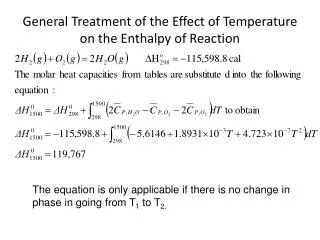
Download
1 / 13
140 likes | 310 Vues
Effect of Liraglutide Treatment on Inflammation and Oxidative Stress in Young and Old APP/PS1 and WT Mice. Supervisors: Dr. Christian Holscher Dr. Kerry Hunter Geisa Nogueira Salles B00627190. Introduction. Substantial anti-oxidant and anti- inflammatory effects in endothelial cells.

E N D
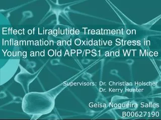
Effect of Liraglutide Treatment on Inflammation and Oxidative Stress in Young and Old APP/PS1 and WT Mice Supervisors: Dr. Christian Holscher Dr. Kerry Hunter Geisa Nogueira Salles B00627190
Introduction Substantial anti-oxidant and anti- inflammatory effects in endothelial cells • Analogue of GLP-1 (glucagon-like peptide-1), hormone released in response to nutrients which potentiates insulin secretion and reduce blood glucose levels Range of neuroprotective properties Liraglutide Treatment with insulin has been shown to improve brain function in humans (Okereke, O. I. et al., 2008) Approved for the treatment of diabetes as an agent that promote insulin secretion (Lovshin and Drucker, 2009)
Oxidative damage to DNA may play a role in normal aging and neurodegenerative diseases (Mecocci, P. et al, 2004) Mechanisms associated to oxidative stress and free-radical reactions have a crucial role in the pathogenesis of AD, specifically, in the formation of plaques and tangles (Lyras L, et al. 1997) 8-OxoguanineCommon DNA lesion caused by reactive oxygen species, associated with neurodegeneration Oxidative Stress in AD Brain Inflammation in AD • Causes degeneration, disease progression and declines cognition IBA-1(ionized calcium-binding associated protein) Macrophage/microglia marker The number of microglia within neuritic plaques is increased β-amyloid Microglia Inflammatory Response ACTIVATES INITIALIZES
Methods 8 OxoGuanine Staining IBA-1 Staining Sections of hipoccampus of these groups were cut in Cryostat • Immunohistological experiment done with 1º and 2º ab • Chromogen was used to colour the sections • Pictures were taken in Zeiss® Axio Scope A1 (cortex for IBA-1 and dentate for 8OxoG) • Pictures were analysed with ImageJ® • Statistical analysis was done with Prism®, using One-way ANOVA • Wild type Saline • Wild type Liraglutide • APP/PS1 Saline • APP/PS1 Liraglutide 9 and 15 month old female (n=6 for each group) treated for 3 weeks
Aims • Compare the effects of Liraglutide in 9 and 15 month old APP/PS1 and WT mice • Determine if Liraglutide is able to interfere and reduce brain inflammation and oxidative stress in APP/PS1 AD mouse model compared to WT mice
Results APP/SI1 Saline APP/SI1 Liraglutide WT Saline WT Liraglutide % IBA-1 expression in mouse cortex of WT and APP/PS1 female mice (n=6 for each group) treated with Liraglutide (25nMol/Kg bw) or Saline (0.9% w,v NaCl) as control for 3 weeks. Values are the mean ± S.E.M *** p<0.001 compared to APP/PS1 Saline; ΔΔΔ p<0.001 compared to APP/PS1 Liraglutide
Results APP/SI1 Saline APP/SI1 Liraglutide WT Saline WT Liraglutide % IBA-1 expression in mouse cortex of WT and APP/PS1 female mice (n=6 for each group) treated with Liraglutide (25nMol/Kg bw) or Saline (0.9% w,v NaCl) as control for 3 weeks. Values are the mean ± S.E.M *** p<0.001 compared to APP/PS1 Saline; ΔΔΔ p<0.001 compared to APP/PS1 Liraglutide
Results APP/SI1 Saline APP/SI1 Liraglutide WT Saline WT Liraglutide % 8OxoGunine expression in mouse cortex of WT and APP/PS1 female mice (n=6 for each group) treated with Liraglutide (25nMol/Kg bw) or Saline (0.9% w,v NaCl) as control for 3 weeks. Values are the mean ± S.E.M *** p<0.001 compared to APP/PS1 Saline.
Results APP/SI1 Saline APP/SI1 Liraglutide WT Saline WT Liraglutide % 8 OxoGuanine expression in mouse cortex of WT and APP/PS1 female mice (n=6 for each group) treated with Liraglutide (25nMol/Kg bw) or Saline (0.9% w,v NaCl) as control for 3 weeks. Values are the mean ± S.E.M * p<0.05 and ** p<0.01 compared to APP/PS1 Saline.
Discussion IBA-1 9 month old IBA-1 15 month old • No significant difference for IBA-1 between WT and APP/PS1 • at 9 and 15 month old. • The microglia activation was not reduced with the treatment, • since the inflammatory marker IBA-1 was not reduced. M1 macrophage: Produce pro inflammatory cytokines, TNFgama, IL-6, ILI2 Activated microglia not only exert neuroprotective effects but might also be detrimental for the survival of neuronal tissue (Luo XG et al, 2010) M2 macrophage: Reduce inflammatory + adaptative TH1 response, producing anti inflammatory factors
Discussion 8OxoGuanine 9 month old 8OxoGuanine 15 month old • The oxidative stress presented significant reduction with • the Liraglutide treatment in APP/S1 for both groups • Liraglutide has considerable anti-oxidant effect in the brain Liraglutide treatment can reduce free radicals associated with typical lesions found in the brains of AD patients, therefore preventing: damage to DNA protein oxidation lipid peroxidation (Christen, Y, 2000)
Conclusion • Liraglutide may be effective in preventing neurodegenerative processes in AD patients, since it prevent oxidative stress in the brain in young and old mice • For the microglia activation, the high expression of IBA-1 may be related to an overexpression of M2 macrophages, which produce anti-inflammatory factors REFERENCES Christen, Y. (2000).Oxidative stress and Alzheimer disease. Am J Clin Nutr 2000 71: 621s Karran, E., Mercken, C., De Strooper, B. (2011). The amyloid cascade hypothesis for Alzheimer’s disease: an appraisal for the development of therapeutics. Nature Rev. Drug Discov., 10, 698-712. Lovell MA, Gabbita SP, Markesbery WR (1999) Increased DNA oxidation and decreased levels of repair products in Alzheimer’s disease ventricular CSF. J Neurochem 72:771–776 Lovshin, J. A., & Drucker, D. J. (2009). Incretin-based therapies for type 2 diabetes mellitus. Nature Reviews Endocrinology, 5(5), 262–269. Luo X.G., Ding Q, Chen D. (2010). Microglia in the aging brain: relevance to neurodegeneration. Molecular Neurodegeneration, 5. Lyras L, Cairns NJ, Jenner A, Jenner P, Halliwell B (1997) An assessment of oxidative damage to proteins, lipids, and DNA in brain from patients with Alzheimer’s disease. J Neurochem 68:2061–2069 Mecocci P, MacGarvey U, Beal MF (1994) Oxidative damage to mitochondrial DNA is increased in Alzheimer’s disease. Ann Neurol 36:747–751 Okereke, O. I. et al. (2008). A profile of impaired insulin degradation in relation to late-life cognitive decline: a preliminary investigation. Int. J. Geriatr. Psychiatry 24, 177–182. Small, D. H., Cappai, R. (2006). Alois Alzheimer and Alzheimer’s disease: a centennial perspective, Journal of Neurochemistry, 99, 708-710. Wimo, A., Prince, M. (2010). World Alzheimer Report 2009: the global economic impact of dementia. Alzheimer’s Disease International.
References • Christen, Y. (2000). Oxidative stress and Alzheimer disease. Am J Clin Nutr 2000 71: 621s • Karran, E., Mercken, C., De Strooper, B. 2011. The amyloid cascade hypothesis for Alzheimer’s disease: an appraisal for the development of therapeutics. Nature Rev. Drug Discov., 10, 698-712. • Lovell MA, Gabbita SP, Markesbery WR (1999) Increased DNA oxidation and decreased levels of repair products in Alzheimer’s disease ventricular CSF. J Neurochem 72:771–776 • Lovshin, J. A., & Drucker, D. J. (2009). Incretin-based therapies for type 2 diabetes mellitus. Nature Reviews Endocrinology, 5(5), 262–269. • Luo XG, Ding JQ, Chen SD. Microglia in the aging brain: relevance to neurodegeneration. Molecular Neurodegeneration. 2010;5 • Lyras L, Cairns NJ, Jenner A, Jenner P, Halliwell B (1997) An assessment of oxidative damage to proteins, lipids, and DNA in brain from patients with Alzheimer’s disease. J Neurochem 68:2061–2069 • Mecocci P, MacGarvey U, Beal MF (1994) Oxidative damage to mitochondrial DNA is increased in Alzheimer’s disease. Ann Neurol 36:747–751 • Okereke, O. I. et al. A profile of impaired insulin degradation in relation to late-life cognitive decline: a preliminary investigation. Int. J. Geriatr. Psychiatry 24, 177–182 (2008). • Small, D. H., Cappai, R. 2006. Alois Alzheimer and Alzheimer’s disease: a centennial perspective, Journal of Neurochemistry, 99, 708-710. • Wimo, A., Prince, M. 2010. World Alzheimer Report 2009: the global economic impact of dementia. Alzheimer’s Disease International. • http://link.springer.com/content/pdf/10.1007%2Fs004010100418 • http://ajcn.nutrition.org/content/71/2/621s.full • Acessed in 14/04/2013