
X-ray Fluorescence and Speciation Analysis of Selenium in A. bisulcatus Taproots and Nodules
This study utilizes µXRF mapping and XANES spectroscopy to investigate the distribution and speciation of selenium (Se) in the taproot and lateral roots of A. bisulcatus. X-ray fluorescence mapping reveals the spatial arrangement of Se (in red), calcium (Ca in green), and iron (in blue) in cross-sections, while speciation analyses at designated locations elucidate the chemical forms of Se within the plant structures. Findings contribute to understanding the roles of Se in plant physiology and potential applications in agriculture and environmental studies.

X-ray Fluorescence and Speciation Analysis of Selenium in A. bisulcatus Taproots and Nodules
E N D
Presentation Transcript
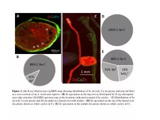
SeCaFe o o A D o o 100% C-Se-C o o 600mm o E C B 46% C-Se-C 89% C-Se-C SeCaZn 23% SeO32- 31% Se0 11% Se0 Figure 1. (A) X-ray fluorescence (µXRF) map showing distribution of Se (in red), Ca (in green) and iron (in blue) in a cross-section of an A. bisulcatus taproot. (B) Se speciation in the tap root as determined by X-ray absorption near edge structure (XANES) spectroscopy at the locations indicated in panel A by circles. (C) Distribution of Se (in red), Ca (in green) and Zn (in pink) in a lateral root with nodule. (D) Sespeciation in the tip of the lateral root (locations shown as white circles in C). (E) Sespeciation in the nodule (locations shown as white circles in C). o o 1 mm