Download
1 / 29
330 likes | 1.13k Vues
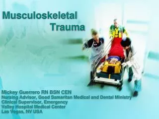
Musculoskeletal Trauma. Mickey Guerrero RN BSN CEN Nursing Advisor, Good Samaritan Medical and Dental Ministry Clinical Supervisor, Emergency Valley Hospital Medical Center Las Vegas, NV USA. Objectives. Identify common mechanisms of injury

E N D
Musculoskeletal Trauma Mickey Guerrero RN BSN CEN Nursing Advisor, Good Samaritan Medical and Dental Ministry Clinical Supervisor, Emergency Valley Hospital Medical Center Las Vegas, NV USA
Objectives • Identify common mechanisms of injury • Discuss the nursing assessment of the patient with musculoskeletal injury • Plan appropriate nursing interventions for the patient with musculoskeletal injury • Evaluate effectiveness of interventions • Understand basic principles of splinting
Physics of Injury • Newton’s first law of motion: An object in motion tends to stay in motion, unless acted upon by an opposing force • Law of conservation of energy: Energy cannot be created or destroyed, only transferred • When a moving body is acted on by an outside force and changes its motion, Kinetic energy must change to some other form of energy • If the moving object is a human and the energy transfer occurs too rapidly, the result is TRAUMA
Physics? Why Is That Important? • History and Mechanism of injury can help predict extent of injuries and prompt assessment for further injuries • The more force involved in the trauma, the more injuries suspected • High energy equals high injury • Consider assessing for the following depending on injury and history: • Hypovolemia, crush syndrome, compartment syndromes, hyperkalemia, sepsis, rhabdomyolisis and fat embolism
Primary Assessment • Begins with assessing airway, breathing, and circulation • After ensuring that no life threatening injury has been untreated, the nurse assess and stabilizes any extremity injuries • Assess for: • Swelling and deformity • Contusion, abrasion, laceration, puncture wound • Bruising • Crepitus • Point tenderness • Neurovascular: distal pulses, color, movement, sensation, temperature, capillary refill, and pain
Before Immobilization • Open fractures should stabilized, and bleeding controlled • Open fractures with obvious bone protrusion or deep laceration should be rinsed with sterile saline (preferably) to remove contamination and then covered with a dry non- adhering sterile dressing • To control bleeding apply pressure directly to injury site, edges of wound, or adjacent pressure point • A tourniquet can be considered on open limb wounds
Immobilization • Should be accomplished as soon as possible to minimize further damage and reduce pain. • A splint should include the area above and below the injury. • Three basic types of splints: • Hard splints such as padded board, cardboard, plaster, and fiberglass. • Soft splints such as pillows. • Traction splints which reduce angulation and provide support.
Skull & Facial Trauma Basilar skull fracture >> • S/S Raccoons eyes from intraorbital bleeding • Combative behavior is common. <<< Battle sign occurs 12 – 24 hours after injury. Blood is collecting behind the tympanic membrane.
Facial Trauma • Palpate before edema and hematomas obscure bony landmarks, stand above head and compare symmetry • Very vascular area, can involve much bleeding • Control bleeding with direct pressure whenever possible • Nasal fracture most common • Assess for open airway • Avoid head tilt method, consider NPA unless midface or nasal fractures are present • Protect cervical spine, assume injury until proven otherwise • Perform brief neurologic exam like GCS and pupil response • Head injuries are a major cause of traumatic death, and cause long term disability
Neck and SpineA Team Centered Approach Maintain patient on backboard and in C-collar until cleared by physician • Removal is team approach. Most important job is maintaining C-spine alignment. Maintain gentle but firm stabilization of neck. Log roll patient away from physician. • Patient in supine position is at risk for aspiration. • Consider airway management early, before edema can compromise breathing.
Types of Fractures Oblique • Twisting force Spiral • Twisting force Comminuted • Severe direct trauma. Avulsed • Forceful contraction of muscle mass. Impacted • Severe trauma causes fractured bone ends to jam together. Greenstick • Partial fracture, common in children because bones are still soft.
Upper Torso Clavicle Fracture • Cannot raise arm. Apply figure 8 support. Sling and swathe if elderly. Apply ice pack intermittently Shoulder fracture • Most frequent in falls with elderly • If non- displaced, only require sling and swathe, or shoulder immobilizer • Truly complicates activities of daily living Upper arm fracture • Casted with splint from axilla around elbow and back to shoulder. Sling applied. Ice for pain and swelling
Upper Extremities Forearm fractures • Usually result from fall • Interventions are splint and sling • The entire arm should be supported • Cast with the elbow at 90 degrees Wrist fractures • Require elevationand splinting Metacarpal and finger fractures • Also called “boxers fracture” are common during striking injuries • Remove rings before swelling increases • Encourage elevation • Buddy tape
Shoulder Dislocation Anterior - Often occurs from falling on extended arm. Posterior – Deformity is difficult to see. Signs and symptoms • Dislocations produce severe pain, joint deformity, point tenderness, and inability to move affected joint. Interventions • X ray before relocation, unless neurovascular compromised. • Analgesia and sedation should be given before reduction. • After reduction place sling and swathe with immobilizer to prevent subluxation. • Remember to assess distal pulses, color, movement, sensation, temperature, and capillary refill before and after relocation. • Obtain after reduction radiology to confirm placement.
Pelvic Fractures Causes • Vehicular trauma, particularly pedestrians, and falls from heights. Highly vascular area • Suspect hemorrhagic shock with tachycardia and hypotension. Interventions • High flow O2, serial vital signs, two large bore IV’s for volume replacement. Prepare for potential blood loss and replacement of 1500 to 4000ml. • Foley catheter NOT recommended. • Immobilize spine and legs. • Flexing knees may decrease pain.
Pelvic Fractures Classification • Stable • Pelvic ring fracture in one place • Stable to palpation • No abnormal deformity • Unstable • Pelvicring fracture in more than one place • Unstable to palpation
Hip Fractures Causes • Falls most common, especially in elderly • Trauma with driver in MVC or motorcycle Signs and symptoms • Pain in the groin, hip, knee, severe pain with movement, and immobility • Trochanteric fractures have increased shortening of extremity, and a greater degree of external rotation Interventions • Early immobilization with Bucks traction or surgical intervention is often necessary • Pain control • Foley placement no longer considered • Place wedge between knees when log rolling
Hip Dislocation Causes • Major trauma while leg is extended, high impact to knee, falls, and crush injuries Signs and symptoms • Pain in hip and knee • Posterior dislocation- hip flexed, adducted, and internally rotated • Anterior dislocation- hip flexed, abducted, and externally rotated Interventions • Check neurovascular status distally: color, movement, sensation, temperature • Splinting is done in presenting position, or position of comfort • Avascular Necrosis of femoral neck may occur if hip is not relocated within 6 hours • Foley is no longer recommended • After joint relocation, patient will begin long period of bed rest with traction
Femur Fracture Signs and symptoms • Severe pain, unable to bear weight • Severe muscle spasms cause leg to shorten, and can move bone ends, causing further injury and pain • Crepitus occurs over fracture site as bone pieces move Interventions • Traction splint. (Long air splint, or other closed foot splints not recommended.) • Two large bore IV’s • Frequent vital signs • Pain control • Prepare for traction or surgery • Assess extremity color, temp, equal pulses
Knee Fracture & Dislocation Signs and symptoms • Knee pain, inability to bend or straighten knee, swelling and tenderness • Patellar fracture has obvious deformity Interventions • Long leg splint, knee immobilizer, or securing one leg to the other • Mid-thigh to mid-calf • Can be made from cardboard or blanket with tape or fabric to hold in place • Treatment is rest, ice, elevation • Knee dislocations require immediate reduction to prevent complications
Tibia and Fibula Fracture Signs and symptoms • Tenderness, swelling, deformity, and crepitus • Many tib/ fib fractures are open Interventions • Open and closed should be splinted as they are found • Realignment should NOT be attempted unless it is open and neurovascular compromise is suspected • Closed fractures are at high risk for compartment syndrome • Rest, ice, elevation “Toes above the nose”
Ankle Injuries Involve distal tibia, distal fibula, or talus. Can be caused by trauma- direct or indirect, and torsion. Signs and symptoms • Pain and inability to bear weight • Swelling and deformity Interventions • Anticipate reduction on closed injury, and walking cast placement. • Potential for surgery and pinning • Monitor neurovascular status: color, temperature, sensation, and capillary refill • Treatment is rest, ice, elevation
Foot, Heel, &Toe Injuries Interventions • Compression dressing for foot • Buddy tape toes • Instruct to wear hard soled wooden or open toe shoes • Crutches may be needed if unable to bear weight • Complications are rare.
Compartment Syndrome • Dangerous swelling, threat to limb circulation • Frequent assessment of neurovascular status and swelling, report circulatory changes immediately!!! • Muscle compartment is sliced open to relieve pressure • Tissues bulge and the result is a massive surgical wound that takes months to heal • This patient is at high risk for infection and amputation • Compartment Syndrome and amputation are a worst case scenario • Anticipate pain needs!!
Amputation Traumatic event for patient • Physically and emotionally Control active bleeding -Amputations may benefit from a tourniquet Remove gross debris Elevate and splint the stump Prepare patient for surgery Place amputated part on ice bath: do not freeze • Wrap in saline soaked gauze • Place in plastic bag then • Place bag on ice or ice water mixture Administer medications for pain, antibiotics for infection
Crush Injuries Life-threatening and difficult to treat • Cellular destruction • Damage to vessels Result from • prolonged entrapment • Crushing blow Concerns • Hemorrhage and fluid loss • Destruction of muscle • Infection • Compartment syndrome • Rhabdomyolysis Nursing Interventions • Administer intravenous fluids to increase urinary output and excrete myoglobin • Elevate the injured extremity • Cleanse open wounds • Reassess urinary output • Monitor motor and sensory function
Points to remember • Clean open wounds. • Cover with sterile non-adhering dressing. • Anticipate pain needs. • Patient at high risk for infection. • Frequent neurovascular assessment of distal pulses, color, movement, sensation, temperature. • Anticipate fluid and blood replacement. - Immobilize early. - Report any changes to physician.
Questions? Contact information Mickey Guerrero email: guerrero6@mac.com Skype: mickey.1972 Facebook: Mickey Guerrero
References • Howard, P. K., Steinmann, R. A., & Sheehy, S. B. (2010). Sheehy's emergency nursing: principles and practice. (6th ed.). St. Louis, Mo.: Mosby Elsevier. • Tintinalli, J. E., & Stapczynski, J. S. (2011). Tintinalli's emergency medicine: a comprehensive study guide (7th ed.). New York: McGraw-Hill. • TNCC: trauma nursing core course (5th ed.). (2000). Park Ridge, Ill.: Emergency Nurses Association.