Download
1 / 32
330 likes | 1.23k Vues
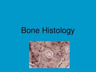
The Histology of Bone and Cartilage. Articular cartilage. Spongy bone. marrow. Compact bone (location of haversian systems). Microscopic Anatomy of Bone. Figure 5.3. Microscopic Anatomy of Bone. Lacunae Cavities containing bone cells (osteocytes) Arranged in concentric rings Lamellae

E N D
Articular cartilage Spongy bone marrow Compact bone (location of haversian systems)
Microscopic Anatomy of Bone Figure 5.3
Microscopic Anatomy of Bone • Lacunae • Cavities containing bone cells (osteocytes) • Arranged in concentric rings • Lamellae • Rings around the central canal • Sites of lacunae Figure 5.3 Slide 5.11a
Microscopic Anatomy of Bone • Canaliculi • Tiny canals • Radiate from the central canal to lacunae • Form a transport system Figure 5.3 Slide 5.11b
T.S. through compact bone x100 Note the numerous ‘Haversian Systems’
Dark spots are called ‘lacunae’ and would contain osteocytes in living bone Central canal containing an artery, vein, lymph vessel and nerves.
What happens when excessive bending force compresses the bone…! compression force
The bone’s good compressive strength was not enough to withstand this particular tackle…!
The matrix of bone is a mixture of organic (collagen) and inorganic (calcium phosphate) 90% of bone is matrix, with the remaining 10% made of osteocytes.
Label the following: A B C Don’t confuse this picture with the Liver Lobule!
Compact bone Compression forces at the junction… Spongy bone
‘snow-boarder’s wrist!’ The fracture occurs at the junction between compact and spongy bone.
CARTILAGE Recognisable by cells called chondrocytes …in small cavities called lacunae surrounded by a matrix of embedded glycoproteins and collagen fibres,
chondrocytes A lacunae B matrix C
Bone contains cells called __________embedded in a matrix. • The organic part of the matrix contains ___________? • Osteoblasts secrete a pre-cursor to this called _________? • The inorganic part contains ________________? • What is the location of blood vessels in compact bone? • Which way do haversian systems run within long bones? • What are the cells that make up cartilage called? • What are they embedded in? www.nyerrn.com
The Vertebral Column • Vertebrae separated by intervertebral discs • The spine has a double curvature • Each vertebrae is given a name according to its location Figure 5.14
Structure of a Typical Vertebrae Figure 5.16
Synovial fluid ligament muscle cartilage Synovial membrane