Download
1 / 11
120 likes | 368 Vues
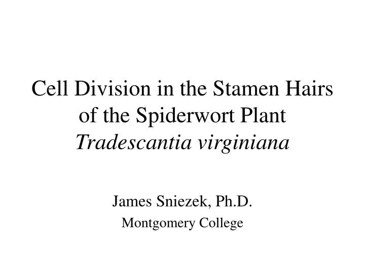
Cell Division in the Stamen Hairs of the Spiderwort Plant Tradescantia virginiana. James Sniezek, Ph.D. Montgomery College. Purpose. Document cell division in a representative plant cell Increase microscopy skills

E N D
Cell Division in the Stamen Hairs of the Spiderwort Plant Tradescantia virginiana James Sniezek, Ph.D. Montgomery College
Purpose • Document cell division in a representative plant cell • Increase microscopy skills • Develop materials to enhance teaching of mitosis and cytokinesis in the classroom
Materials & Methods • Isolate a young bud from T. virginiana • Dissect bud on a clean microscope slide isolating the stamens from the flower • Keep cells moist in 15mm HEPES/ 15mm KCl • Isolate stamen hairs from stamen • Cover with 22mm2 coverslip, view with microscope, keep moist
Materials & Methods • View at 400x magnification with a Zeiss IM35 microscope equipped with differential interference contrast and a Panasonic time-lapse video system. • Images were also captured using NIH image 1.6 and stored on an apple xxxxx
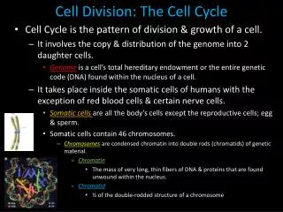
Mitosis is described as nuclear division without a reduction in chromosome number Each new nucleus (daughter nucleus) will have the same quantity of DNA as the parental nucleus If cytoplasmic division (cytokinesis) also takes place then two identical daughter cells are formed Mitotic events can be categorized into discrete stages based on what is happening to structures within the cell Stages include: Prophase Prometaphase Metaphase Anaphase Telophase What’s going on?
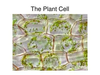
Cell structures to follow • Structures of the nucleus • Nuclear envelope (defines the nucleus): this structure will breakdown during prometaphase and be reformed during anaphase. • Chromatid (strand of DNA associated with chromosomes): these structures will condense to become apparent in cells undergoing nuclear division (prophase) and will elongate following their separation (anaphase). • Centromere (region of chromosome containing the kineotchore that joins sister chromatids): during anaphase the centromeric area cleaves allowing the sister chromatids to be separated. • Structures of the cytoplasm • Spindle MTOC: responsible for the coordinated assembly of microtubules used in forming the spindle apparatus. • Spindle fibers: microtubules that grow toward the chromatids; some attach to the kinetochore.
Interphase Cell • Cell performs normal function • Three subphases: • G1: cell duplicates most organelles • S: quantity of DNA in the cell is doubled. Each chromosome has a pair of sister chromatids connected by kinetochores • G2: chemical components stockpiled • Nucleolus present
Prophase(Including Prometaphase) Three things visibly occur • Chromosomes condense resulting in visible chromatids • Spindle MTOC’s producing spindle fibers • Nuclear envelope breaks down
Metaphase • Chromosomes are moved by growing spindle fibers to the equator of the cell (metaphase plate) Region of centromere chromatid
Anaphase • Kinetochore splits into two • Spindle fibers shorten from kinetochore end separating sister chromatids and pulling them toward the poles
Telophase • Chromatids lengthen becoming less apparent • Nuclear membrane reforms around each region of chromosomal material • Cytokinesis occurs as cell plate is formed • Nucleolus reforms