Download
1 / 72
980 likes | 2.27k Vues
Bone: Fracture Patterns Bone Healing Fracture Management. PTP 521 Musculoskeletal Diseases and Disorders. Objectives. Describe forces involved in fracturing a bone Describe common bone fracture patterns Differentiate between fracture patterns by discussing forces involved

E N D
Bone: Fracture Patterns Bone Healing Fracture Management PTP 521 Musculoskeletal Diseases and Disorders
Objectives Describe forces involved in fracturing a bone Describe common bone fracture patterns Differentiate between fracture patterns by discussing forces involved Discuss three different types of fracture classification Discuss bone healing Discuss fracture management
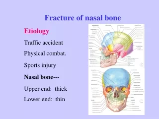
Fractures: • Break in the continuity of the bone • Physical Therapy Practice Pattern 4G: Impaired Joint Mobility, Muscle Performance and Range of Motion Associated With Fracture
Risk Factors or Consequences of Pathology – Guide to PT Practice Bone demineralization Hormonal changes Medications Menopause Nutritional deficiency Prolonged immobilization or non-weight bearing state Trauma
Causes: • 1. External forces: • 2. Internal forces: nutrition • 3. Pathology: at risk to fracture other bones.
Clinical Manifestations: Signs and Symptoms of a Fracture ** Most Common Manifestations 1. Signs: • **Deformity of the bone • **Edema • **Ecchymosis: is the swelling, purple-blue color. • **Loss of general function and/or mobility • WB? 2. Symptoms: • **Pain • **Point tenderness over fracture site • Increase symptoms with vibration or tapping
Body Structure Dysfunction, Activity Limitations or Participation Limitations – PT Guide to Practice Inability to access community Limited Range of Motion: body structure/dysfunction. Muscle weakness from immobilization: body structure/dysfunction Pain with functional movements and activities: activity limitation
Fracture Descriptions Site of fracture Extent of fracture Fracture Alignment Direction of Fracture Lines Special Features of Fractures Associated Abnormalities Special Types of Fractures
Site of the Fracture A. Diaphyseal : B. Metaphyseal: C. Epiphyseal: D. Intra-articular: E. Fracture-Dislocation:
Trauma Registry System of Fracture Classification A. Long bones 1) intra-articular 2) extra-articular B. Flat bones 1) intra-articular 2) body or extra-articular
Extent of the Fracture A. Complete: fracture which breaks both cortex -harder to heal, poorer alignment. B. Incomplete: fracture which breaks only one cortex
Fracture Alignment: Fracture patterns predicted by the external force and bones inherent characteristics. Load: application of force, type of load can determine fracture type. Common Loads: 1) Tension: pull 2) Compression: WB 3) Bending: a little flexibility in bone. 4) Torsion:
Fracture Alignment • Position: Relationship of fragments to their normal anatomical structure • Need 2 x-rays at 90 degrees to each other to see a fracture. • Alignment: Distal segment in relation to proximal segment • Relationship of the longitudinal axis of one fragment to the other • Apposition: The placement of two bone segments in close proximity
Position: Fracture Alignment • Non-displaced: Fracture segments are in good alignment and don’t require any intervention for alignment • Displaced: Loss of apposition between segments
Relationship of Fracture Fragments to Each Other B. Displaced: fracture whose ends are separated due to: • Force of the injury: high (more likely to displace) vs low • Gravity • Pull of muscles attached to the bone: muscle spasm (often after injury) so may pull bone out of alignment (often rotation)
Types of Displacement a. Shifted: Apposition is present • Bones usually unite • Fractures surfaces may not be in contact with one another in one plane • Medial, Lateral, Anterior, Posterior, Superior or Inferior to each other
Fracture in a 2 year old • Where is the fracture? • Femur • What direction is it shifted? • Lateral Displacement • The image on the right was taken at the time of injury, with the leg in a fiberglass cast.
b. Angulated: tilted, if not corrected, could lead to a deformity in the limb
Types of Displacement c. Rotated: bone looks straight but one limb is rotated about its longitudinal axis
Types of Displacement • Distracted: ends are separated and pull apart from each other • Issue is with soft tissue going between the ends, then can’t heal.
Types of Displacement e. Overriding: • muscle spasms with the injury • the bones are pulled past each other • shortens the bone
Direction of Fracture Lines Reference: longitudinal axis of the bone Irregular shaped bones are referenced by the cortex of the bone
Naming of Fracture Patterns • Depends upon • Load – what type • Direction of fracture lines
Basic Patterns 1)Transverse Fracture • Fracture is perpendicular to the long axis of the bone • Load: bending force • Low energy • Stable, fracture fragments usually remain in place
Basic Patterns 2) Longitudinal: fracture line runs parallel to the long axis of the bone • Load: may be a repetitive stress or an extension of an oblique fracture • Risk: Tibial Longitudinal Fractures: runners, jumpers (basketball players) and old women
Basic Patterns 3. Oblique fracture: diagonal break across the bone • Load: axial compression, bending and torsion force • Moderate energy
Basic Patterns 4) Spiral Fracture: fracture is jagged, pointed ends, can have soft tissue damage • Load: torsion force • Low energy
oblique Spiral
Comminuted Butterfly fracture • Load: axial compression and bending force • Moderate energy • Butterfly Fractures occur on side of concavity of the fracture
Comminuted Segmental Fracture • Comminuted fracture, bone in more than two parts • 3-100 or more
Special Features of Fractures • Impacted: fractured bone is driven into itself, shortens the bone • Impactions • Load: axial compression force • Variable energy is required • Fracture lines can be indistinct as the fracture ends are jammed together
Basic Patterns Avulsion Fractures http://sacs.vetmed.ufl.edu/notes/CROSS/response.htm
Associated Abnormalities Fracture dislocation of the ankle, BoneandSpine.com Subluxations: Partial dislocation, still within the confines of the joint capsule Dislocations: bones are completely disarticulated, outside the joint capsule
Special Types of Fractures Stress fractures Pathologic fractures Periprosthetic fractures Bone graft fractures
Types of Incomplete Fractures seen in Children • Greenstick Fracture • Plastic Bowing: low load, prolonged stress. • Torus Fracture (Buckle): load is really high so folds in on itself.
Salter-Harris Classification of Children’s Epiphyseal Fractures • Type I: separation of epiphyseal plate • Type II: separation of epiphyseal plate plus metaphyseal wedge fracture • Type III: separation of epiphyseal plate plus epiphyseal wedge fracture
Type IV: metaphyseal and eipiphyseal fracture fragment • Type V: impaction fracture of epiphyseal plate and adjacent surfaces
Salter Harris Classifications • Rang’s type VI: • Involves the epiphysis at the outside periphery of the bone – perichondrial ring • May cause an osseous bridge between the metaphysis and epiphysis.
Ogden’s VII • Tip of the epiphysis • Articular surface is involved • Increases risk of OA later in life.
Ogden VIII • Metaphysis fracture that disrupts the blood flow to the epiphyseal plate
Ogden IX • Avulsion fracture of the periosteum which can influence the growth plate and/or vascular supply to the epiphyseal plate
Closed Fracture Descriptors • Closed (simple): fracture that doesn’t break the skin • Tscherne Classification • Grade 0: no soft tissue damage, indirect forces, torsion fractures • Grade 1: superficial abrasion or contusion caused by fragment pressure from within. Mild to moderate fracture severity
Grade 2: deep contaminated abrasion, local skin or muscle contusion from direct trauma • Grade 3: skin extensively contused, crushed muscle, severe muscle damage, vascular injury, compartment syndromes are common
Open Fracture Descriptors • Open fractures (compound): open wound is present • Gustillo Classification • Type I: wound is less than 1 cm long, low energy trauma, minimal soft tissue damage, no signs of crush injury • Type II: wound is more than 1 cm long, slight to moderate crush injury, no extensive soft tissue damage, flap or avulsion • Type III: extensive wound and soft tissue damage, greater degree of fracture comminution and instability, high degree of contamination
Fracture Complications A. Uncomplicated: fracture that heals uneventfully B. Complicated: 1) nonunion: failure of the bone fragments to unite 2) malunion: healing of a fracture occurs, but a deformity results 3) delayed union: healing at a fracture site that progresses too slowly compared to the norms 4) posttraumatic OA: altered joint mechanics due to an intra-articular fracture or malunion
Complications • Open fracture complications – Infections • Compartment Syndrome: 5 P’s • Pain, Pallor, Paresthesia, Paralysis, Pulselessness • Nerve Injury • Arterial Injury • Infection • Complex Regional Pain Syndrome • Limb Length Discrepancy
Life Threatening Complications (Open or Closed) Fat embolism: stroke Hemorrhage Pulmonary embolism: air bubbles Gas Gangrene: when muscle tissue dies, so amputation is only way to stop it. Tetanus: bacteria that first sign is slok-jaw, rigidity of all muscle.
Cortical Bone Healing After a Fracture: Inflammatory Stage 1) Fracture gap is < 10uM • Osteoclasts are present only when the bony ends die back • If gap is small (< 10 uM) – no bone death will take place and the healing occurs with Haversian remodeling • No osteoclasts are present • If gap is greater than 10uM, Haversian remodeling can’t occur and the regular healing takes place
2) Fracture gap is > 10uM • 0 – 3 days after a fracture • Mesenchymal cells arrive, produce a fibrous tissue that envelops both ends of the fracture site • Macrophages are present to clean out the debris • Hematoma is beginning to be absorbed • Fibrin clot develops between the fracture ends
Proliferative Stage: 1) Soft Callus Formation: • Cells: osteogenic and chondrogenic cells, osteoclasts • Clinical Union: callus has united at the fracture site • No movement at fracture site • Fracture is NOT normal in strength • Callus: contains fibroblasts, blood vessels, cartilage and new bone • Mechanical characteristics change