Download
1 / 1
30 likes | 325 Vues
Leukemia Cutis. Histopathology. Acknowledgements. Conclusion. Differential Diagnosis. Results. Introduction. Patient Description. Intervention. Leukemia Cutis: A Diagnostic Symptom Seen at Presentation and Relapse in Acute Myeloid Leukemia

E N D
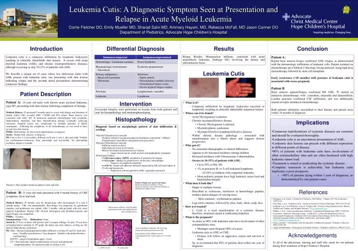
Leukemia Cutis Histopathology Acknowledgements Conclusion Differential Diagnosis Results Introduction Patient Description Intervention Leukemia Cutis: A Diagnostic Symptom Seen at Presentation and Relapse in Acute Myeloid Leukemia Corrie Fletcher DO,Emily Mueller MD, SharadSalviMD, AmmaryHayani, MD, Rebecca McFall, MD Jason Canner DO Department of Pediatrics, Advocate Hope Children’s Hospital Leukemia cutis is a cutaneous infiltration by neoplastic leukocytes resulting in clinically identifiable skin lesions. It occurs with acute myeloid leukemia (AML) and chronic myeloproliferative diseases, although occurring in only 10-15% of patients with AML. We describe aunique set of cases where two adolescent males with AML present with leukemia cutis: one presenting with skin lesions indicating relapse and the seconds initial presentation demonstrating cutaneous findings. Biopsy Results: Mononuclear infiltrate, consistent with acute monoblastic leukemia, (Subtype M5) involving the dermis and subcutaneous tissue . Patient A: Repeat bone marrow biopsy confirmed AML relapse, as demonstrated with the dermatologic infiltration of leukemic cells. Patient restarted on chemotherapy per Children’s Oncology Group protocol, using high dose chemotherapy followed by stem cell transplant. Early recurrence (<18 months) with presence of leukemia cutis is associated with worse prognosis. Patient B Bone marrow aspirate/biopsy confirmed M4 AML. Pt started on induction chemotherapy, with cytarabine, etoposide and daunorubicin. Lumbar puncture confirmed CSF infiltration, and was additionally started on triple intrathecal chemotherapy. Both patients ultimately succumbed to their disease and passed away within 10 months of diagnosis. • Patient A: 14 year old male with known acute myeloid leukemia, type M5, presenting with skin lesions following completion of therapy. • Medical History: Pt presented 9 months prior to event with fatigue and shortness of breath. Initial CBC revealed WBC 174,000 and 85% blasts. Bone marrow was consistent with AML, M5. Pt underwent induction chemotherapy with cytarabine, daunorubicin, etoposide, and then intensification therapy eventually achieving remission. Hospital course was complicated by multiple episodes of febrile neutropenia. Following the completion of all chemotherapy, pt was noted to have several new skin lesions. • PMHx: Mild asthma. No previous hospitalizations or surgeries. • Sochx: Denies etoh, tobacco, or drug use. • PE: Four to five 1-2 cm nodules, diffusely on R arm, L arm, L shin and scalp. Nodules are erythematous/violaceous, firm, non-tender and non-mobile. No surrounding erythema, edema or warmth. • What is it? • Cutaneousinfiltration by neoplastic leukocytes (myeloid or lymphoid), resulting in clinically identifiable cutaneouslesions. • Where can it be found? • Acute Myelogenous Leukemia • Chronic myeloproliferative disease • Chronic Myelogenous Leukemia • Myelodysplastic syndromes • Myeloproliferative/lymphoproliferative diseases • Within chronic disease pathology - associated with transformation into a blastic phase and suggests disease progression. • Who gets it? • No consistent demographic or clinical differences. • Appears to be increased incidence among children. • Increased incidence with Chromosome 8 abnormalities. • Occurs in 10-15% of patients with AML. • Up to 50% in M4, M5. • 1% in precursor B- or T-cell leukemia/lymphoma. • 25-30% of children with congenital leukemia. • Most pediatric patients have high leukemic tumor load and hepatosplenomegaly. • What does it look like? • Single or multiple lesions. • Described as violaceous, red-brown or hemorrhagic papules, nodules and/or plaques of varying sizes: • Most common - erythematous papules. • Legs most common, followed by arms, back, chest, scalp, face. • How is it treated? • L. Cutis is a local manifestation of a systemic disease; therefore, treatment aimed at eradicating leukemia. • What is the prognosis? • As many as 90% with leukemia cutis have involvement of other extramedullarysites: • Meninges most frequent (40% of cases). • Leukemia cutis in AML or CML: • Disease will follow an aggressive course and survival is short. • Su, et al estimated that 88% of patients died within one year of diagnosis. Excisional biopsies were performed on lesions from both patients and sent for histopathology and immunophenotyping. Implications • Cutaneous manifestations of systemic diseases are common and should be evaluated thoroughly. • Leukemia cutis is an uncommon presentation of AML. • Leukemic skin lesions can present with different expression at different points of disease. • 90% of patients with leukemia cutis have involvement of other extramedullary sites and are often burdened with high leukemic tumor load. • Treatment is aimed at eradicating the systemic disease. • Complete remission is achievable, but leukemia cutis implicates a poor prognosis. • >80% of patients dying within 1 year of diagnosis, as demonstrated by our patient cases. • Diagnosis based on morphologic pattern of skin infiltration & cytology • Myeloid (Granulocytic) sarcoma • Nodular with perivascular-periadnexal distribution (interstitial + diffuse) • Predominately myeloblasts and granulocytic precursors • Myeloid (Monoblastic) sarcoma • Infiltrate similar to above • Subcutaneous tissue involvement • Blastic chromatin and promonocytic features • Large cleaved nuclei, small nucleoli and amphophilic cytoplasm • Histological Stains: • (+)Myeloperoxidase(MPO) -neoplasms of granulocytic lineage • (+)Lysozyme- Marker for granulocytes, monocytes, macrophages • Negative in lymphoid neoplasms • (+)CD 56 - neural cell adhesion molecule expressed by NK cells and subset of • T cells and monocytes • Expression well recognized in AML, especially monocytic Patient A Right arm; punch skin biopsy Patchy mononuclear infiltrate of deep adenexalstructures with confluent infiltrate in subcutaneous adipose tissue Patient A: Skin nodules found on patient’s arms and shin References Patient B: 15 year old male presented with 4 month history of URI symptoms, fever and rash. Medical History: Pt initially seen by dermatology after development of a rash 4 months earlier. CBC was unremarkable. Skin biopsy was diagnostic for granulomaannulare, and prednisone was started. After 3 months, rash persisted with new onset gingival hyperplasia. Repeat CBC showed neutropenia and thrombocytopenia and repeat biopsy was completed. PMHx: + Eczema Immunizations: UTD. Medications: None Social hx: Pt lives at home with parents and 3 younger siblings. No pets. Pt was born in England and is currently in 10th grade. He denies any etoh, tobacco, or drug use. He follows Halal dietary restrictions PE: Skin—Raised, hyperpigmented nodules diffusely covering UE and LE, back and torso. Variable sizes between 1-2cm diameter. Nodules with defined borders and blanching. HEENT—gingival hyperplasia upper >lower LN—Post-auricular, anterior and posterior cervical, and submental lymphadenopathy. No supraclavicular or axillary LAD • Kleigman et al. Nelson’s Textbook of Pediatrics 18th Edition. “Chapter 495, The Leukemias.” 2120-2122. • Cho-Vega J, et al. “Leukemia cutis.” American Journal of Clinical Pathology. 2008;129:130-142. • Goldman et al. Cecil Medicine, 23rd ed. “Chapter 466: Urticaria, Drug Hypersensitivity Rashes, Nodules and Tumors, and Atrophic Diseases.” www.mdconsult.com • Shipley J, et al. “Acute myeologenous leukemia.” Experimental Hematology 2009;37:649-658. • Curesearch: Children’s Oncology Group. “AAML0531: A Phase III Randomized Trial of Gentuzumab Ozogamicin Combined with Conventional Chemotherapy for De Novo Acute Myeloid Leukemia (AML) in Children, Adolescents, and Young Adults.” www.childrensonologygroup.org • Pearce and Sills. “Consultation with the Specialist: Childhood Leukemia” Pediatrics in Review. 2005;26:96-104. • Segel and Halterman. “Neutropenia in Pediatric Practice.” Pediatrics in Review. 2008;29:12-24 . Subcutaneous infiltrate of polymorphic mononuclear cells with wrinkled nuclei (low and high power) Acute monoblastic leukemia, M5 involving dermis and subcutaneous tissue of skin Karyotyping of skin infiltrate showed rearrangement in the 11q23 region in 173 of 200 cells To all of the physicians, nursing and staff who cared for our patients during their treatment at Hope Children’s Hospital. L: CD45 (LCA) R: Leder stain