Download
1 / 16
160 likes | 314 Vues
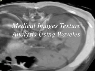
Binning Strategies for Tissue Texture Extraction in DICOM Images CTI Students: Bikash Bhattacharyya, Kriti Jauhar Advisors: Dr. Daniela Raicu, Dr. Jacob Furst Submitted To: RSNA Conference ‘05, Chicago , IL.

E N D
Binning Strategies for Tissue Texture Extraction in DICOM Images CTI Students: Bikash Bhattacharyya, Kriti Jauhar Advisors: Dr. Daniela Raicu, Dr. Jacob Furst Submitted To: RSNA Conference ‘05, Chicago, IL
Binning Definition: Putting gray-levels into bins for image compression e.g. 1,2,3,4 gray levels in Bin 1 5,6,7,8 gray levels in Bin 2DICOM Images – 12 Bit - 4096 intensitiesTexture Feature Calculation – All intensities - SLOWBinning allows for additional flexibility to trade off large intensity ranges against computational speed Why Binning ? COMPUTATION PERFORMANCE
Linear Binning Linear Binning - Bins of equal size 256 bins for DICOM images produces bin ranges [0..15] , [16..31] ,…[4081..4096] Quick and Efficient approach Pre -processing step for Harlick texture feature calculation Promising results for classification of tissues based on Haralick texture features
Disadvantages of Linear Binning • Soft tissues with similar intensities may end up in the same bin with linear binning Soft tissues misclassification Accuracy of Liver and Spleen not very high Computed Tomography images contain low number of pixel in the range [1500 – 4096] Non-Linear Binning – Is it possible to improve accuracy of soft tissues?
Analysis of Linear Binning (contd.) EXAMPLES Spleen Liver
Two Approaches of Non-LinearBinning Non Linear Binning based on K-Means Clustering 256 Clusters – Compare results of 256 linear-bins Distance Measure – Euclidean Clusters of Gray Level Ranges Gray Level ranges form Non-linear Bins Clipped Binning based on visual inspection of gray levels Range [0, 856] mapped to Bin 1 Range [1368 , 4096] mapped to Bin 258 Range [856, 1368] mapped to 256 linear bins e.g. 856 to 858 gray levels in Bin 123
Non-linear Binning using K-Means Process Flow
K-Means • K =256 • 141 Dimensions/Images • 4096 points/Gray Levels • Initial Points /Random Centroids • Similarity Metric = Euclidean Distance Issues • 263 Unique Gray Levels Identified • Multiple Gray Levels – Identified in one Cluster e.g. Cluster 14 has gray levels from 462 to 884 Cluster 14 also has gray levels from 1540 to 1542
Conclusion • Non-Linear Binning with K-Means gave us the best overall results ( 86.35%) • Results for Liver and Spleen improved from 73.80% to 91.03% for liver and 70.50% to 74.58% for spleen • Clipped Binning performed poorly on testing set with overall sensitivity of only 68.85% • Results with K-Means improved over Linear Binning
Future Work • Experimenting with bins other than 256 such as: 64, 128 etc. • Exploring other similarity measures such as: Jeffrey Divergence, Mahalanobis Distance etc. • Testing other classification algorithms besides decision trees, such as: Neural Networks, Bayesian Networks, Logistic Regression etc.
References • [1]M. Kalinin, D. S. Raicu, J. D. Furst, D. S. Channin,, " A Classification Approach for Anatomical Regions Segmentation", The IEEE International Conference on Image Processing (ICIP), September 11-14, 2005. (submitted) • [2] D. Channin, D. S. Raicu, J. D. Furst, D. H. Xu, L. Lilly, C. Limpsangsri, "Classification of Tissues in Computed Tomography using Decision Trees", RSNA, DECEMBER, 2004. • [3] R.M. Haralick, K. Shanmugam, and I. Dinstein, “Textural Features for Image Classification”, IEEE Trans. on Systems,Man, and Cybernetics, vol. Smc-3, no.6, pp. 610-621, 1973. • [4] N. M. Nasrabadi and R. A. King., “Image Coding Using Vector Quantization: A review” , IEEE Transaction on Communications, 36(8):957-971, August 1988. [5] Wei-Ying Ma and B. S. Manjunath, “A Texture Thesaurus for Browsing Large Aerial Photographs”, Journal Of The American Society For Information Science, 49(7):633–648, 1998. • [6] Dongqing Chen, Lihong Li, and Zhengrong Liang, “A Self-adaptive Vector Quantization Algorithm for MR Image Segmentation” , ISMRM,1999. • [7] Qixiang Ye2 Wen Gao Wei Zeng1, “Color Image Segmentation Using Density-Based Clustering”, ICASSP, 2003, Presentation.
References • [8] Martin Ester, Hans-Peter Kriegel, Jorg Sander, XiaoWei Xu, “A Density-Based Algorithm for Discovering Spatial Databases With Noise,” Proceedings of 2nd International Conference on Knowledge Discovery and Data Mining, 1996. • [9] Texture Classification of Normal Tissues in Computed Tomography D. Xu, J. Lee, D.S. Raicu, J. D. Furst, D. Channin, The 2005 Annual Meeting of the Society for Computer Applications in Radiology, Orlando, Florida, June 2-5, 2005. • [10] N. B. Karayiannis, "Soft learning vector quantization and clustering algorithms based on ordered weighted aggregation operators," IEEE Transactions on Neural Networks, vol. 11, no. 5, pp. 1093-1105, 2000. • [11] N. Papamarkos and B. Gatos, "A new approach for multithreshold selection", Computer Vision, Graphics, and Image Processing-Graphical Models and Image Processing, Vol. 56, No. 5, pp. 357-370, Sept. 1994 • [12] A. Atsalakis, N. Kroupis , D. Soudris, and N. Papamarkos, "A window-based color quantization technique and its architecture implementation", ICIP2002, Rochester, USA. • [13] N. Papamarkos, A. Atsalakis and C. Strouthopoulos, "Adaptive Color Reduction", IEEE Trans. on Systems, Man, and Cybernetics-Part B, Vol. 32, No. 1, Feb. 2002.