Download
1 / 11
120 likes | 346 Vues
Analysis of Thin Microkeratome Flap Architecture Using Fourier-Domain OCT. Karolinne Maia Rocha, M.D., Ph.D J. Bradley Randleman, M.D. R. Doyle Stulting , M.D., Ph.D. The authors have no financial interest. Emory University. Purpose.

E N D
Analysis of Thin Microkeratome Flap Architecture Using Fourier-Domain OCT Karolinne Maia Rocha, M.D., Ph.D J. Bradley Randleman, M.D. R. Doyle Stulting, M.D., Ph.D. The authors have no financial interest Emory University
Purpose To assess the corneal architecture and the reproducibility of laser in situ keratomileusis (LASIK) flap thickness created by a mechanical microkeratome using a Fourier-domain optical coherence tomography (OCT).
Methods • Prospective study • 58 LASIK flaps (Amadeus II - Ziemer,140µm head, 9.0 and 9.5mm suction rings, ML7090CLB - Med-Logics blades) • Intraoperative ultrasound (US) pachymetryflap thickness measurement • Fourier-domain OCT (Optovue RTVue-100) flap morphology evaluation at 2 weeks postoperatively
Methods Fourier-domain OCT Analysis The flap thickness was assessed twice at : • 10 points across the central 6-mm of the corneal apex (horizontal and vertical meridians) • 1.5-mm from the edge of the flap superior and inferiorly
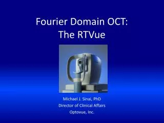
Time Domain vs Fourier Domain • RTVue device (Optovue) • 26,000 A scans/second • depth resolution of 5 µm • transverse resolution of 15 µm • Scan beam wavelength: 840±10 nm • Exposure power at pupil 750 W • Cornea–Anterior Module (CAM) • additional software on the device, • The CAM L lens allows 6 x 6-mm • scans of the cornea • Visante OCT 1000 (Carl Zeiss) • 2000 A scans/second • depth resolution of 18 µm • transverse resolution of 60 µm • Scan beam wavelength: 1310 nm • Exposure power at pupil 5 W • The scan range is 3 mm in depth to 10 mm in transverse direction.
Results • IntraoperativeUS pachymetry= 107.2±14µm • Postoperative OCT = 111.7±11µm • No statistically significant difference (p=0.07) • The OCT measurements showed a planar flap • SD across the flap = 4.9µm, • SD for the 6-mm central measurements = 11µm • SD range across the flap= 2 to 9µm
Conclusion • The Amadeus IImicrokeratome with Med-Logics blades created regular, planar flaps as demonstrated by the Fourier-domain OCT. • Central flap thickness measured by intraoperativeUS pachymetrywas comparable to that measured 2 weeks postoperative by OCT.
Thank you karolinnemaia@gmail.com