Download
1 / 9
100 likes | 396 Vues
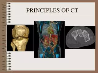
Magnetic Resonance Imaging & Positron Emission Tomography. AUBREY OVERSON. OUTLINE. MRI ~A bit of physics of the system ~image reconstruction. PET SCAN ~positron emission ~making of isotopes ~detector. COMPARISON. Positron Emission Tomography. Simultaneously & collinear

E N D
Magnetic Resonance Imaging & Positron Emission Tomography AUBREY OVERSON
OUTLINE MRI ~A bit of physics of the system ~image reconstruction PET SCAN ~positron emission ~making of isotopes ~detector COMPARISON
Positron Emission Tomography • Simultaneously & collinear • produces two photons • with energy 511 KeV (mec2) • travel nearly 180º back to back • this is measured by a coincidence detector
CYCLOTRON Also has better radionuclides with a low atomic numbers (11C, 13N, 15O, 18F) which have a higher affinity of the human body as well as relatively short half lives… 11C=20.34min; 13N=9.96 min; 15O=2.05 min; 18F =110min;
Scintillation Camera collimator crystal photomultiplier coincidence circuit
IMAGE RECONSTRUCTION 2D projection 3D Image Filtered Back-projection (FBP). MRI contrast works by altering the local magnetic field in the tissue being examined. Normal and abnormal tissue will respond differently to this slight alteration, giving us differing signals. These varied signals are transferred to the images, allowing us to visualize many different types of tissue abnormalities and disease processes better than we could without the contrast. http://www.seas.gwu.edu/~gogo/movies/homer3d.mov
Magnetic Resonance Imaging(MRI) Larmor Frequency
More MRI details When the RF pulse is turned off, the hydrogen protons begin return to their natural alignment within the magnetic field and release their excess stored energy. When they do this, they give off a signal that the coil now picks up and sends to the computer system. What the system receives is mathematical data that is converted, through the use of a Fourier transform, into a picture that we can put on film. That is the "imaging" part of MRI.
COMPARISONS PET 500 machines in US Metabolic processes Advantage of false color More sensitivity and resolution than SPECT or CT $2000-4000 dollars per scan MRI Very common does not use ionizing radiation is a comfort to many patients, ability to image in any plane. It gives images of brain tumors, abscesses, swelling, bleeding, nerve damage, and other disorders that increase the fluid content of tissues.