Download
1 / 37
370 likes | 509 Vues
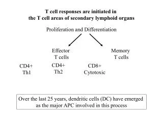
In spite of the considerable body of evidence that CD28 provides a critical costimulatory signal for T cell responses, not all T cell-mediated responses are CD28-dependent. Kunding T.M. et al. Immunity 5:41, 1996. VSV does not replicate extensively in vivo, in contrast to LCMV.

E N D
In spite of the considerable body of evidence that CD28 provides a critical costimulatory signal for T cell responses, not all T cell-mediated responses are CD28-dependent.
VSV does not replicate extensively in vivo, in contrast to LCMV. Continued presence of signal 1 alone, either through prolonged viral replication or repeated injection of peptide, generates a functional T cell response in vivo in absence of CD28. The duration of TCR stimulation determines the costimulatory requirement of T cells.
Schweitzer, A.N. and Sharpe A.H. J. Immunol. 61:2762, 1998. 4 days T cells + WT APC pulsed with peptide T cells
London C.A. et al. J. Immunol. 164:265, 2000. TCR Trsg T cells + WT APC pulsed with peptide 4 days T cells Transferred into mice 6-7 months Memory T cells Stimulated in vitro with WT APC B7 KO APC
In the presence of strong or prolonged signal 1, T cells don’t need costimulation. Costimulation is not required for effector and memory T cells. Now, In the presence of strong signal 1, T cells don’t need CD28-mediated costimulation. CD28-mediated costimulation is not required for effector and memory T cells. These responses may involve additional costimulatory pathways.
B7h/ICOS costimulatory pathway A new molecule with structural characteristic similar to the B7 molecules was identify in 1999, and was named B7h (B7-related protein 1; also GL-50 or B7RP-1 or ICOS-L). B7h does not bind to CD28 or CTLA-4, but bind to ICOS (inducible costimulatory molecule). ICOS shares 30-40% sequence similarity with CD28 and CTLA-4. ICOS expression: ICOS is not constitutively expressed on naïve T cells but is induced on CD4+ and CD8+ T cells following stimulation through the TCR and is further enhanced by CD28-mediated costimulation. FIGURE 2. Expression of ICOS on activated T cells. Dissociated splenocytes from wild-type or B7-1/2-/- 129/SvS4Jae mice were incubated with anti-CD3, anti-CD3 and CD28, or no Ab. The thick line shows ICOS expression on T cells from wild-type splenocyte cultures, the dotted line shows ICOS expression on T cells from B7-1/2-/- splenocyte cultures, and the thin line represents a negative staining control (rat IgG-FITC). McAdam A.J. et al. J. Immunol. 165:5035, 2000.
ICOS is expressed on memory T cells (CD45RO+) and on T cells in germinal centers. b, Localization of ICOS+ cells in the apical light zone of germinal centres, as determined from histochemical stains of frozen human tonsillar sections with monoclonal antibody F44 using the APAAP technique30. LZ, light zone; DZ, dark zone; MZ, mantle zone (original magnification 69). Hutloff A. et al. Nature 397:263, 1999. More highly expressed on Th2 than Th1 clones. ICOS expression decreases during the differentiation of Th0 cells to Th1. McAdam A.J. et al. J. Immunol. 165:5035, 2000. Coyle A.J. et al. Immunity 13:95, 2000.
B7h expression • Strongly expressed in the B-cell area of the lymph nodes and spleen and in germinal centers. • Induced on DCs and monocytes • Induced on non-lymphoid cells (kidney, lungs and testis) by inflammatory stimuli The expression of B7RP-1 is strong in the lymph nodes from both normal and sensitized mice (Fig. 2a), particularly in the cortex (a B-cell area) and in both primary and secondary follicles. In other lymphoid tissues, B7RP-1 expression is primarily localized in B-cell areas of the spleen, the follicles in the Peyer's patches and the medulla of the thymus (Fig. 2b). Yoshinaga S.K. et al. Nature 402:827, 1999.
Activation of naive and recently activated CD4+Tcells 2- Second activation OVA peptide + + CD4+ OVA-specific TcR Tg APC CTLA-4 Ig ICOS Ig hIg Ig ICOS Ig CTLA-4 Cytokine production (ELISA) Experimental procedure Naive T cells Recently activated T cells 1- First activation of CD4+ T cells with APC-peptide + + OVA peptide CD4+ OVA-specific TcR Tg APC hIg Proliferation IL-2 production (ELISA) Coyle A.J. et al.(2000). Immunity, 13: 95-105.
Activation of naive and recently activated CD4+Tcells in vitro ICOS-Ig ICOS-Ig CTLA-4Ig CTLA-4Ig hIg • ICOS is not implicated in the activation of naive CD4+ T cells and production of IL-2. • ICOS pathway is important for cytokine production from recently activated CD4+ T cells.
Cytokine production by fully differentiated Th1 and Th2 effectors cells Experimental procedure Threerounds of repetitive stimulations with OVA peptide CTLA-4 Ig + + ICOS Ig CD4+ T cells APC hIg Cytokine production (ELISA) Th1 Th2 Coyle A.J. et al.(2000). Immunity, 13: 95-105.
Cytokine production by Th1 and Th2 effectors cells Coyle A.J. et al.(2000). Immunity, 13: 95-105. • B7RP-1/ ICOS pathway contributes to the cytokine production from Th2 but not Th1 cells in vitro.
ICOS +/+ ICOS +/- ICOS -/- T cell-dependent Ab production and germinal center formation in ICOS -/- miceExperimental procedure Primary Immunization with KLH/ CFA Antibody response (ELISA) GC formation (Immunohistochemistry) Second Immunization with KLH/ CFA Cytokine production (Immunostaining and cytofluorimetry) Tafuri A. et al (2001). Nature, 409: 105-109.
Antibody response and germinal center formation in ICOS -/- mice ICOS +/+ ICOS +/- ICOS -/- Tafuri A. et al (2001). Nature, 409: 105-109. ICOS is required for antibody responses and GC formation.
ICOS +/+ T cells ICOS -/- T cells Cytokine production after restimulation in vitro Tafuri A. et al (2001). Nature, 409: 105-109. ICOS is required for Th2 differentiation.
Additional in vivo evidences that Th2 responses are primarily regulated by B7h/ICOS pathway Mucosal model of inflammation in the lung (Th2-mediated) Gonzalo JA et al (2001). Nature Immunol, 2: 597-604. Blocking B7h/ICOS during priming: no effect Blocking B7/CD28 during priming: inflammation Blocking B7h/ICOS during effector phase: inflammation Blocking B7/CD28 during effector phase: no effect EAE (Th1-mediated disease) EAE in ICOS deficient animal : Exacerbation of the disease Dong C et al (2001). Nature, 409: 97-101. Blocking B7h/ICOS during antigen priming : Increased disease severity Rottman JB et al (2001). Nature Immunol, 2: 605-611. augmentation of Th1 polarization by inhibition of Th2 polarization
In vivo evidences that B7h/ICOS pathway also regulatesTh1 responses Th1 cells express lower levels of ICOS compared to Th2 cells. In vitro studies have indicated that ICOS is not relevant in Th1 polarized T cells. However, in vivo studies indicate that expression of ICOS by Th1 cells appears to be relevant EAE (Th1-mediated disease) Rottman JB et al (2001). Nature Immunol, 2: 605-611. Blocking B7h/ICOS during antigen priming : Increased disease severity augmentation of Th1 polarization by inhibition of Th2 polarization Blocking B7h/ICOS during effector phase: Limits disease progression by inhibiting Th1 functions (IFNg). Th1-mediated cardiac allograft rejection: Ozkaaaynak E et al (2001). Nature Immunol, 2: 591-596. Blocking ICOS prolongs allograft survival by preventing both acute and chronic rejection. Thus, these results show that ICOS is also important in regulating Th1 responses in vivo.
Role of B7h/ICOS in CD8 T cell responses • B7h/ICOS interactions are not relevant in CD8 T cell responses to LCMV • Kopf M et al (2000)J. Exp. Med 192: 53-62. • New studies indicate that ICOS may be relevant for effective CD8 T cell mediated • anti-tumor responses: • Wallin JJ et al (2001) J. Immunol. 167: 132-139. • Liu X et al (2001)J. Exp. Med 194: 1339-1348. • In both studies, expression of B7h on tumor cells results in efficient CD8 • mediated tumor rejection. (Delayed kinetic compared with rejection of • B7.2-expressing tumors).
PD-L1, PD-L2 and their receptor PD-1 • PD-L1 (programmed death 1 ligand),PD-L2 (programmed death 2 ligand) and do notbind to CD28, ICOS, or CTLA-4, but bind to PD-1 (programmed death 1 molecule). • PD-1 shows 24 % of sequence similarity to CD28 and CTLA-4. • An ITIM motif (Immunoreceptor Tyrosine-based Inhibitory Motif) is present in the cytoplasmic tail of PD-1. The ITIM motif is known to be present in receptors that have an inhibitory function on lymphocyte responses.
Expression patterns of PD-L1, PD-L2 - PD-L1 and PD-L2 are expressed on DCs and monocytes. - They are constitutively expressed on several non-lymphoid tissues. Dong H. et al (1999). Nature Med, 5(12): 1365-1369 Latchman et al. (2001). Nature Immunol., 2(3):261-268 Coyle A.J. et al (2001). Nature Immunology, 2(3): 203-209
Expression pattern of PD-1 • PD-1 is constitutively expressed on T cells following activation. • (Chambers, C. (2001) Trends Immunol., 22(4): 217-223). • C57Bl/6 PD-1 -/- mice develop lupus-like arthritis and glomerulonephritis with an increase in serum IgG3 and B cell proliferation.(Nishimura, H. el al (1999). Immunity, 11: 141-151). • BALB/c PD-1 -/- develop an autoimmune dilated cardiomyopathy, with IgG deposites on cardiomyocytes and an increase of IgG in blood.(Nishimura, H. el al (2001). Science, 291: 319-322). • CTLA-4 has a stronger negative effect than PD-1 on T cell activation. All Ig isotypes are elevated in CTLA-4 -/- mice independently of the genetic background. The lymphoproliferation is more severe, and the onset of the phenotype and the lethality occur faster in CTLA-4 -/- mice. (Chambers, C. (2001) Trends Immunol., 22(4): 217-223).
PD-L1-Ig hIg CD3 CD3 CD4+ T cells T cell proliferation and cytokine production after PD1- PD-L1 engagement Experimental procedure + Proliferation Cytokine production (ELISA) Freeman et al (2000). J. Exp. Med, 192 (7): 1027-1034.
T cell proliferation and cytokine production after PD-1/ PD-L1 engagement Freeman et al (2000). J. Exp. Med, 192 (7): 1027-1034 PD-L1/ PD-1 interactions can inhibit TcR-mediated proliferation and cytokine production.
Proliferation and cytokine production after PD-L2/ PD-1 engagement Experimental procedure OVA peptide + APC First activation OVA-specific TcR Tg CD4+ T cells CHO.I-Ad CHO.I-Ad.PD-L2 OVA peptide Second activation + Proliferation Cytokine production (ELISA) Latchman et al. (2001). Nature Immunol., 2(3):261-268
Cytokine production after PD-L2/ PD-1 engagement Latchman et al. (2001). Nature Immunol., 2(3):261-268 PD-L2/ PD-1 interactions can inhibit TcR-mediated proliferation and cytokine production.
Contradicting Results as to the function of the PD-L2/PD-L1/PD-1 pathway
Costimulation of T cells by PD-L2 Tseng, SY et al. (2001). J. Exp. Med., 193 (7): 839-845. • PD-L2 costimulates : • a T cell proliferative response to a greater level than B7.1. • mainly CD4+ T cells. • greater levels of IFN- than B7-1, and fails to promote IL-4 or IL-10 production. • PD-L2 appears to drive Th1 responses.
Contradicting results • T cell proliferation and IL-10 secretion increased with PD-L1 stimulation. Dong et al (1999). Nature Medecine, 5(12): 1365-1369). Tamura et al (2001). Blood, 97(6): 1809-1816. • It is unclear whether the PD-L1/L2 costimulation reported by these groups is PD-1 dependent or whether it could be mediated by an alternative receptor for PD-L1: Parallel of CD28/ CTLA-4 receptors- B7.1/ B7.2 ligands. Freeman et al (2000). J. Exp. Med, 192 (7): 1027-1034
B7-H3 B7-H3 • Expression pattern: • B7-H3 (B7 homolog 3)binds to an unknown receptor on activated T cells that is distinct from CD28, CTLA-4, ICOS or PD-1. Ctrl serum • B7-H3 is an inducible molecule on the surface of DCs and monocytes. • Not restricted to lymphoid tissue but also observed in heart, kidney, testis, colon and human tumor lines Transient expression of the unknown receptor of B7-H3 on activated T cells. B7-H3 Ig Ctrl Ig Chapoval et al (2001). Nature Immunology, 2(3): 269-274
Costimulating T cell responses by B7-H3Experimental procedure ImmobilizedB7-H3 Ig B7-1 Ig control Ig Melanoma cell line transfected with B7-H3 or CT vector Coated CD3 + T cells T cells CTL activity Proliferation Cytokine production Chapoval et al (2001). Nature Immunology, 2(3): 269-274
Costimulating T cell responses by B7-H3 Chapoval et al (2001). Nature Immunology, 2(3): 269-274 • B7-H3: • increases T cell proliferation but is less potent than B7.1. • enhances proliferation of both CD4+ and CD8+ T cells. • B7-H3- transfected cells induces CTL activity. • selectively enhances IFN production with modest effects on TNFa and IL-8
Resting T cell CD28 CTLA-4 Primed T cell PD-1 Primed T cell Summary + Initiation of T cell activation. APC B7.1 B7.2 - Inhibition of T cell activation APC (B) + T cell activation in 2nd responses, differentiation in Th2 B7RP-1 ICOS Primed T cell DC, M PD-L1 - Negative regulation of T cell activation. PD-L2 + ? Th1 responses (INF ). DC, M + Th1 (INF) and CTLs. B7-H3 ? Primed T cell
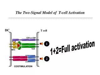
Conclusion • Regulation of T cell activation by costimulation is more complex than originally envisioned. • None of the newly discovered pathways appears to be completely redundant with CD28 in terms of naïve T cell activation but rather regulate the fate of primed and memory T cells. • The spirit of the two-signal model of T-cell activation remains intact, but a second activation step process called « step two of signal 2» can be add to this model. Coyle A.J. et al (2001). Nature Immunology, 2(3): 203-209
Complementation in functions: ICOS/B7h: Th2 ?/B7-H3: Th1 and CTL • Spatial differences in expression: The novel B7 family members have a broad distribution in non-lymphoid tissues and are therefor uniquely position to regulate antigen-specific T cell functions at sites of inflammation.