Hydrocephalus
Hydrocephalus. Presaented by : Faisal Hussain . Majid Ahmed. Lecture Objective :. Definition Epidemiology. Anatomy and Physiology Classification . Pathogenesis . Etiology . Clinical feature . Diagnosis Management. Definition and Epidemiology :. Definition :

Hydrocephalus
E N D
Presentation Transcript
Hydrocephalus Presaented by : Faisal Hussain . Majid Ahmed .
Lecture Objective : • Definition • Epidemiology. • Anatomy and Physiology • Classification . • Pathogenesis . • Etiology . • Clinical feature . • Diagnosis • Management .
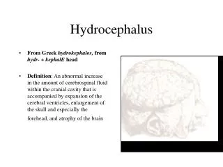
Definition and Epidemiology : • Definition : Hydrocephalus is a disorder in which the cerebral ventricular system contains an excessive amount of cerebrospinal fluid (CSF) and is dilated because of increased pressure. • Epidemilogy: The prevalence of congenital and infantile hydrocephalus has been estimated as 0.48 to 0.81 per 1000 live and still births
Physiology: • CSF Production: site: choroid pluxes Amount : 20ml/h . Rate: 0.1 to 26 ml/h . wich affected by : age and weight Total volume : Range from 50 to 150 ml . CSF produced by active secretion and diffusion. • CSF Absorption : CSF is absorbed into the systemic circulation primarily across the arachnoidvilli into the venous channels of the sagittal sinus
Classification : • Non communicating (obstructive ) The obstruction occurs at theInterventricular foramina, the aqueduct of Sylvius, or the fourth ventricle and its outlets . Note: The proximal area of ventricle system is diliated . • Communicating (non obstructive); due to : 1- decrease absorption : inflammation of the subarachnoid villi . 2- increased secretion .e.g choroid pluxespapilloma
Pathology: • Acute obstruction : 1- causes increased pressure and rapid enlargement of the ventricular system. The frontal and occipital horns of the lateral ventricles enlarge first. Symmetric dilatation of the remainder of the intracerebral CSF-containing spaces follows. 2-Iflattening of the gyri and compression of the sulciagainst the cranium, 3-obliterating the subarachnoid space over the hemispheres. 4-The vascular system is compressed, and the venous pressure in the dural sinuses increases. 5-. contributes to the development of interstitial edema of the periventricular white matter. 6-Another compensatory mechanism that limits expansion of the ventricular system in infants is spreading of the cranial sutures. • chronic hydrocephalus the force of the fluid is distributed over the greater surface area of the enlarged ventricular system
Etiology : • Congenital : A - Neural tube defect : e.gmyelomeningocele has the following 1- obstruction of fourth ventricular outflow 2- flow of CSF through the posterior fossa due to the Chiari malformation 3- aqueductalstenosis . B- Isolated hydrocephalus : aqueductalstenosis in wich this stenosis may due to malformation or inflamation . c- X-linked hydrocephalus : aqueductalstenosis D- CNS malformation : 1- Chiari II portions of the brainstem and cerebellum are displaced caudally into the cervical spinal canal. This obstructs the flow of CSF in the posterior fossa 2- Dandy Walker syndrome :atresia of the foramine of Luschka and Magendie 3- Vein of Galen malformation : compression of the cerebral aqueduct .
Etiology : continued • Congenital continued : E- Intrauterine infection . rubella, cytomegalovirus, toxoplasmosis, and syphilis F- Syndromi Hydrocephalus :13 ,18 ,9 • Acquired : 1- Infection e.g. meningites and encephalities . 2- Tumor : especially posterior fossamedulloblastomas, astrocytomas, and ependymomas. 3- hemorrhage :a- subarachnoid space b- into the ventricular system
Symptoms: • Symptoms in infants • Poor feeding • Irritability • Reduced activity • Vomiting • Symptoms in children • Slowing of mental capacity • Headaches (initially in the morning) that are more significant than in infants because of skull rigidity • Neck pain suggesting tonsillarherniation • Vomiting, more significant in the morning • Blurred vision: This is a consequence of papilledema and later of optic atrophy • Double vision: This is related to unilateral or bilateral sixth nerve palsy • Stunted growth and sexual maturation from third ventricle dilatation: This can lead to obesity and to precocious puberty or delayed onset of puberty.(hypothalmous) • Difficulty in walking secondary to spasticity: This affects the lower limbs preferentially because the periventricular pyramidal tract is stretched by the hydrocephalus. • Drowsiness
Signs : • Children • Papilledema: if the raised ICP is not treated, this can lead to optic atrophy and vision loss. • Failure of upward gaze: This is due to pressure on the tectal plate through the suprapineal recess. The limitation of upward gaze is of supranuclear origin. When the pressure is severe, other elements of the dorsal midbrain syndrome (ie, Parinaud syndrome) may be observed, such as light-near dissociation, convergence-retraction nystagmus, and eyelid retraction (Collier sign). • Macewen sign: A "cracked pot" sound is noted on percussion of the head. • Unsteady gait: This is related to spasticity in the lower extremities. • Large head: Sutures are closed, but chronic increased ICP will lead to progressive macrocephaly. • Unilateral or bilateral sixth nerve palsy is secondary to increased ICP. • Infants • Head enlargement: Head circumference is at or above the 98th percentile for age. • Dysjunction of sutures: This can be seen or palpated. • Dilated scalp veins: The scalp is thin and shiny with easily visible veins. • Tense fontanelle: The anterior fontanelle in infants who are held erect and are not crying may be excessively tense. • Setting-sun sign: In infants, it is characteristic of increased intracranial pressure (ICP). Ocular globes are deviated downward, the upper lids are retracted, and the white sclerae may be visible above the iris. • Increased limb tone: Spasticity preferentially affects the lower limbs.The cause is stretching ofthe periventricular pyramidal tract fibers by hydrocephalus.
Diagnosis : • Serial head measurement . • The diagnosis is confirmed by neuroimaging In a newborn, ultrasonography is the preferred technique due to mobility and has no radition . Infant and children CT and MRI . • A lumbar puncture (LP) should be performed in case of meningities or encephalities .
Differential Diagnosis : • Intracranial Hemorrhage • Intracranial Epidural Abscess • Epidural Hematoma • Subdural Empyema • Subdural Hematoma • Brainstem Gliomas • Meningioma • PseudotumorCerebri: Pediatric Perspective • Pituitary Tumors
Management : • Shunt : RT lateral ventricle to peritoneum . The catheter is connected to a one-way valve system Complication : 1-Infection: Staphylococcus epidermidis , S. aureus, enteric bacteria, diphtheroids, and Streptococcus species. 2- malfunction .
Management : continued • Medical Management : Diuretics . Fibrinolytic therapy . Serial lumbar punctures .