Download
1 / 55
570 likes | 829 Vues
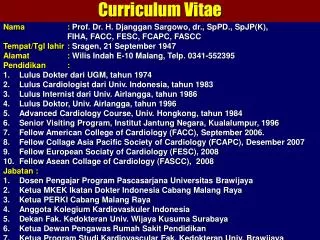
Curriculum Vitae. Nama : Prof. Dr. H. Djanggan Sargowo , dr., SpPD . , SpJP (K), FIHA, FACC , FESC, FCAPC, FASCC Tempat / Tgl lahir : Sragen , 21 September 1947 Alamat : Wilis Indah E-10 Malang, Telp . 0341-552395 Pendidikan : Lulus Dokter dari UGM, tahun 1974

E N D
Curriculum Vitae • Nama :Prof. Dr. H. DjangganSargowo, dr., SpPD.,SpJP(K), • FIHA, FACC, FESC,FCAPC, FASCC • Tempat/Tgllahir :Sragen, 21 September 1947 • Alamat :Wilis Indah E-10 Malang, Telp. 0341-552395 • Pendidikan : • Lulus Dokterdari UGM, tahun 1974 • Lulus Cardiologist dari Univ. Indonesia, tahun 1983 • Lulus Internist dari Univ. Airlangga, tahun 1986 • Lulus Doktor, Univ. Airlangga, tahun 1996 • Advanced Cardiology Course, Univ. Hongkong, tahun 1984 • Senior Visiting Program, InstitutJantung Negara, Kualalumpur, 1996 • Fellow American College of Cardiology (FACC), September 2006. • Fellow Collage Asia Pacific Society of Cardiology (FCAPC), Desember 2007 • Fellow European Sociaty of Cardiology (FESC), 2008 • Fellow Asean Collage of Cardiology (FASCC), 2008 • Jabatan : • DosenPengajar Program PascasarjanaUniversitasBrawijaya • Ketua MKEK IkatanDokter Indonesia Cabang Malang Raya • Ketua PERKI Cabang Malang Raya • AnggotaKolegiumKardiovaskuler Indonesia • DekanFak. Kedokteran Univ. Wijaya Kusuma Surabaya • Ketua Dewan Pengawas Rumah Sakit Pendidikan • Ketua Program Studi Kardiovascular Fak. Kedokteran Univ. Brawijaya
STEM CELL THERAPHY IN CARDIOVASCULAR DISEASES Djanggan Sargowo Surabaya, 12 Mei 2012
O V E R V I E W • What’s the problem? • Hype? • Reality? • Hope? • Conclusions
U N M E T M E D I C A L N E E D • Diabetes • 12.1 million sufferers in US (2002) • Costs $132 billion p.a. (2002) rising to $156 billion p.a. (2010) • Cardiovascular disease • 64.4 million cases in US (2004) • Costs $368.4 billion p.a. (2004) • Parkinson’s Disease • 100,000 sufferers in UK • Treatment costs £600 million p.a. (1998) Paul Rodgers, 2006, Ithaka Lifesciences
DEGENERATIVE DISEASES • Other examples • Alzheimer’s, spinal cord injuries, Amyotrophic Lateral Sclerosis, multiple sclerosis, liver disease etc • Restoration of cell or organ function • Current drugs don’t treat the cause • Organ transplants • Expensive, donor shortages, not applicable to many diseases Paul Rodgers, 2006, Ithaka Lifesciences
Background • Coronary heart disease is the #1 cause of morbidity and mortality in the US. • CHF is the #1 cause of hospitalization for those age > 65 yo. • Annual health care costs related to cardiovascular diseases was ~ $220 billion last year. • Stem cell transplant is a promising and exciting therapy. Orlic D, et al. Nature 2001 2004 American Heart Association Update
Challenge: Knowledge Explosion “We are drowning in information but starved for knowledge.”—Naisbitt, ‘82 Boenjamin Setiawan, dr.,PhD
AVAILABLE STEM CELLS • Bone marrow stem cells (BMSC) • Endothelial progenitor cells (EPC) • Mesenchymal stem cells (MSC) • Skeletal myoblasts (SKM) • Embryonic stem cells (ESC) • Cardiac stem cells (CSC) • Cardiac progenitor cells (isl1+) **Stem cells are capable of self-renewal, transformation into dedicated progenitor cells, and differentiation into specialized progeny Joseph Wu, MD, PhD; Department of Medicine/Cardiology; Department of Radiology/Nuclear Medicine; Email: joewu@stanford.edu
Shinya Yamanaka & James Thomsonpotential Nobel Prize Winners in the Future Shinya Yamanaka,45, working in Japan's Kyoto University with mouse cells, made the iPS breakthrough. He screened 24 candidate proteins before finding four that were able to reprogram adult cells, reverting them to their embryonic state. He and others then showed that these factors are also effective in human cells. Developmental biologist James Thomson,49, of the University of Wisconsinwas the first to identify a slightly different group of factors that do the same . Boenjamin Setiawan, dr.,PhD
VANCOUVER, BRITISH COLUMBIA, MAY 3, 2010 – The March of Dimes awarded Shinya Yamanaka, MD, PhD, (third from left) its 2010 Prize in Developmental Biology at a gala event. Boenjamin Setiawan, dr.,PhD
Induced pluripotent stem cells – the science and technology Chi-Wei Lu, Ph.D. Assistant Professor, RWJMS/UMDNJ Director, Human Embryonic Stem Cell Facility Albert Lasker Basic Medical Research Award, 2009 Boenjamin Setiawan, dr.,PhD
EMBRYONIC STEM CELLS • Immortal • A single cell line is all you need • Precursors of all cell types • Can produce any cell you need • Can divide without limit • Unlimited supply Paul Rodgers, 2006, Ithaka Lifesciences
EMBRYONIC STEM CELLS SURVEY • ES cell therapy is 10-15 years away • Will be used to treat certain diseases • Diabetes, cardiovascular, single gene defects, MS, Alzheimer’s, Parkinson’s, liver disease, spinal cord injury, retinal disease • Schering and Merck are the only pharma companies to say they are interested at this stage Paul Rodgers, 2006, Ithaka Lifesciences
ADULT STEM CELLS • Can be isolated from most tissues • e.g. bone marrow, cord blood, fat, skin etc • Generate cell types of tissue of origin • Produce useful cells for therapies • Plasticity • Can be coaxed into forming cells of completely different tissues • e.g. neurons from blood cells Paul Rodgers, 2006, Ithaka Lifesciences
CLINICAL USE OF ADULT STEM CELLS • Bone marrow transplants • Replace blood cells ablated by cancer therapies • ~ 300 clinical studies on haematopoietic stem cells • Cell expansion is a key issue • Haematopoietic stem cells may be able to restore function to damaged cardiac tissue (3 trials so far) • Neural stem cell trial started in 2006 • Batten’s disease (Stem Cells Inc.) • Developments in China and Korea Paul Rodgers, 2006, Ithaka Lifesciences
INDUCED PLURIPOTENT CELLS AND TRANSPLANTATION THEORY Healthy or diseases adult human or mouse Adult cells (skin fibroblasts) OCT4 SOX2 NANOG Lin28 OCT4 SOX2 KLF4 (Myc) Genetic repair by homo- logous recombination (if necessary) Self renewal iPS cells Differentiation In vitro screening of drug candidates on healthy and diseased cells Tranplan-tation
Cell Sources for Cardiac Repair CSC Endothelial Progenitor Cells Hematopoietic SCs Mesenchymal SCs Hemangioblasts SP cells MAPC Sca-1+ cells Myoblasts SP cells Mesenchymal SCs SPcells Chronic Acute
CARDIAC PROGENITOR CELLS IN THE FETAL AND ADULT HEART Blastocyst Fetal or adult heart ES cells Cardiovascular differentiation Cardic progenitor cells (Kit+, SCA1+, SP or MDR1+) (NKX2 2-5+ or lsl1+) In vitro differentiation Tranplatation In vivo differentiation Vascular smooth muscle cells, endothelial cells or cardiomyocytes Vascular smooth muscle cells, endothelial cells or cardiomyocytes Tissue engineering Mice & other species Tranplatation
TRANSPLANTATION STRATEGY FOR CARDIAC REPAIR Human ES cells Differentiating human ES cells ES-cell-derived cardiomyocytes Adult stem cells from bone marrow Cell injected into the myocardium of imunodeficient mice after myocardial infarction Cardiac progenitor cells Functional analysis by magnetic resonance imaging, ultrasonography or pressure-volume loops Analysis after immunostaining of heart tissue sections
THERAPEUTIC IMPLICATIONS OF CARDIAC PROGENITOR CELLS Human Mouse Pluripotency genes Fetal Adult Blastocyst Skin ES cells iPS cells CARDIAC PROGENITOR CELLS -actinin SMA PECAM Models of heart development Models of disease Drug testing Tissue engineering CARDIAC THERAPHY
Tissue Engineered Myocardium Ischemic heart disease is one of the leading causes of morbidity and mortality in Western societies with 7,100,000 cases of myocardial infarction (MI) reported in 2002 in the United States alone Within 6 years of MI, 22% of men and 46% of women develop CHF MI and CHF will account for $29 billion of medical care costs this year in the US alone Cardiac transplantation remains the best solution, but there is an inadequate supply of donor organs coupled with the need for life-long immunosuppression following transplantation From www.aic.cuhk.edu.hk/web8/Hi%20res/Heart.jpg Boenjamin Setiawan, dr.,PhD
Decrease in EPCs associated with CV disease Endothelial Progenitor Cells Vasculoprotective agents CV risk factors Atherosclerosis Disease Regression? Disease Progression Improvement of endothelial function Enhanced re-endothelialization Reduced plaque size Improved angiogenesis Myocardial infarction Ischemic stroke Erectile dysfunction Renal insufficiency Peripheral artery disease Werner N, Nickenig G. Arterioscler Thromb Vasc Biol. 2006;26:257-66.
CV risk factors Endothelial dysfunction Collaterals Restenosis CV disease EPCs in CV diseases EPCs Pathophysiology Therapeutics Atherosclerosis Heart disease Peripheral vascular disease Courtesy of Arshed A. Quyyumi, MD.
EPC physiology • Originate in bone marrow • Circulate in blood stream • Number and function (proliferation, migration, homing) modulated by age, CV risk factors, and disease • Release stimulated by organ and vascular injury • Participate in vascular repair (collateralization) and re-endothelialization, partly by paracrine effects • Circulating numbers by exercise and drugs (statins and ACE inhibitors) • Independent predictors of endothelial dysfunction and long-term prognosis in patients with CAD Hill JM et al. N Engl J Med. 2003;348:593-600.
Normal vessel Pal-1 Pal-1 EC SMC tPA Atherosclerotic plaque EC SMC Pal-1 Pal-1 tPA Christ et al., Senescent SMCs Increase Vascular Wall PAI-1 Expression
Procoagulant, antifibrinolytic matrix- degrading, leukocyte binding endothelial cell Resting endothelial cell Resting smooth muscle cell IL-1, TNF Collagen Elastin IL-1 TNF Activated matrix-degrading smooth muscle cell Collagenase Gelatinases Elastolytic enzimes Class II MHC Antigen Apoptotic smooth muscle cell IFN TNF T-cell antigen receptor T-lymphocyte
Thrombus Lipid core Adventitia Unstable Coronary Artery Disease (II) Thrombus forms and extends into the lumen
Clinical classification of ACS Acute Coronary Syndrome (ACS) No ST Elevation ST Elevation MI (NSTEMI) MI (STEMI) Unstable Angina Pectoris No Q-wave Q-wave National Heart Foundation of Australia, Cardiac Society of Australia and New Zealand.Med J Aust 2000;173 (suppl):S65–S88
Acute infarction(hours) Infarct expansion (hours to days) Global remodelling (days to months) BACKGROUND ON REMODELLING Improvement of LV remodelling has been associated with improvement in mortality and morbidity outcomes in CHF
MANAJEMEN SKA - Oklusi > 4-6 jam Nekrosis miokard irreversible IMA dengan gel q (IMA-Q) - Reperfusi Menurunkan morbiditas - mortalitas - Fase Pre hospital stage Hospital stage : - IGD - CVCU
CELL-TRANSPLATATION STUDIES IN MODELS OF EXPERIMENTAL MYOCARDIAL INFARCTION
RC Trials using Intracoronary BMC post MI MI size ND ND (n=60) + 2.8% (P=n.s.)* P=n.s. (n=84) ND (n=184) -28% (P=0.03) LEUVEN-AMI (n=67) FINCELL +7.1% (P=0.05) ND (n=80) * 18-months follow-up
Myocardial Homing and Biodistribution of 18F-FDG-labeled BMC 50 to 75 min after Transfer 3D PET 1.3% to 2.6% in infarct (center) Unselected BMC 14% to 39%in infarct (border zone) CD34-enriched BMC Hofmann et al., Circulation 2005; 111: 2198-202
111In uptake in patient after an anterior AMI (cell administration 5 days after acute PCI) Schachinger, V. et al. Circulation 2008;118:1425
Ischemic Cardiomyopathy Therapy Anno 2010: from drugs to cells? • Clinical outcome in ischemic cardiomyopathy patients with LV dysfunction remains unacceptable with combined event rates of 25% despite state-of-the art treatment. • Modest improvement in cardiac function in RCTs of BMC transfer is attributable to: • limited homing, engraftment, and survival of BMCs • lack of cardiac muscle regeneration • (differences in cell infusate)
Conclusions: Cell Therapy for Heart Failure 2010 • Mixed bone marrow-derived progenitor cells have paracrine trophic effects in the dysfunctional ischemic heart. Whether they can affect clinical outcome in patients with large MI, at risk for maladaptive remodeling and heart failure, remains to be determined in prospective RCT. • Identification of cell-specific effects and enhancement of progenitor cell functionality warrant focused trials. • Major breakthrough requires a better understanding of post-natal cardiomyogenesis and strategies to stimulate endoge-nous regeneration, paralleled by effective neovascularisation.
Conclusions: Cell Therapy for the Dysfunctional Heart 2010 • Modest improvement in cardiac function in 6 RCTs of BMC transfer is attributable to: • limited homing, engraftment, and survival of cells • lack of cardiac muscle regeneration • differences in cell infusate • Progenitor cell transfer is best reserved for patients with large MI, at risk for heart failure. • Identification of best cell type, enhancement of cell functionality and stimulation of endogenous cardiac repair warrant focused translational trials. Science Push!
EXPEDITED REVIEW Transplantation of Progenitor Cells and Regeneration Enhancement in Acute Myocardial Infarction Final One-Year Results of the TOPCARE-AMI Trail
Objectives Background Method Result The Transplantation of Progenitor Cells And Regeneration Enhancement in Acute Myocardial Infarction (TOPCARE-AMI) trial investigates both safety, feasibility, and potential effect on parameters of myocardial function of intracoronary infusion of either circulating progenitor cells (CPC) or bone marrow-derived progenitor cells (BMC) in patients with acute myocardial infarction (AMI) A total of 59 patients with AMI were randomly assigned to receive either CPC (n=30) or BMC (n=29) into the infarct artery at 4.9 1.5 days after AMI. Conclusion Intracoronary infusion of progenitor cells (either BMC or CPC) is safe and feasible in patients after AMI successfully revascularized by stent implantation. Both the excellent safety profile and observed favorable effect on LV remodelling, provide the rationale for larger randimized doble-blind trials. (J Am Coll Cardiol 2004;44:1690-9) 2004 by the American Collage of Cardiology Foundation.
TRIAL DESIGN 59 patients with acute MI undergoing successful PCI / sten revascularization - LV angigraphy - BMC Intracoronary infusion of bone-marrow cells; n = 29 CPC Intracoronary infusion of circulating progenitor cells Pregenitor cell theraphy 3 – 7 days Post stent revascularization 4 month clinical follow-up n = 30 n = 29 n = 30 12 months clinical follow-up n = 29 Patients excluded from exploratory analysis n = 1 AMI & death Addational AMI n = 1 < 105 cells received n = 1 n = 27 n = 27 *4 months LV angiography n = 27 n = 18 n = 27 * 4 & 12 months MRI
PROCEDURAL SAFETY OF INTRACORONARY PROGENITOR CELL INFUSION Both cell CPC BMC Groups (n = 30) (n = 29) ( n = 54) Procedural complications* 0 0 0 CRC (mg/dl) Before cell theraphy 2.8±2.2 (2.0) 3.5±2.6 (2.6) 3.1±2.4 (2.3) 24 h after cell therapy 2.6±2.3 (1.8) 3.2±2.0 (2.8) 2.9±2.2 (2.3) 14 d after cell therapy (n=48) 0.65±0.54 (0.5) 1.1±1.3 (0.6) 0.82±0.97 (0.5) 4 months follow-up 0.49±0.38 (0.3) 0.40±0.18(0.3) 0.44±0.30(0.3) Troponin Before cell theraphy 2.3±1.9 (1.7) 2.5±2.1 (1.9) 2.4±2.0 (1.85) 24 h after cell therapy 1.5±1.4 (1.2) 1.9±1.8 (1.5) 1.7±1.6 (1.4) 14 d after cell therapy (n=48) 0.02±0.03 (0.01) 0.03±0.04(0.01) 0.03±0.04 (0.01) Value are expressed as mean ± SD (median). *Thrombosis, embolization, or disection related to cell infusion.
EVENTS-FREE SURVIVAL OF DEATH, RECURRENT MYOCARDIAL INFARCTION, OR TARGET VESSEL REVASCULARIZATION (Kaplan-Meier analysis) 100 90 80 70 60 50 40 30 20 10 0 76 % Event-free survival (%) Death, myocardial infarction, Infarct vessel revasc 0 100 200 300 days number exposed 59 57 57 46 45 45 45 45 to risk