Autonomic Nervous System
Autonomic Nervous System. Dr Richard Tunstall Warwick Medical School GEM 2013. Objectives.

Autonomic Nervous System
E N D
Presentation Transcript
Autonomic Nervous System Dr Richard Tunstall Warwick Medical School GEM 2013
Objectives State and demonstrate the regions of the spinal cord / the brainstem nuclei and the specific spinal and cranial nerves involved sympathetic and parasympathetic nerve outflow from the central nervous system. Explain the location and structure of the sympathetic chain, and outline the range of patterns of synapse of pre- and post ganglionic sympathetic fibres Outline the potential causes and consequences of a loss of sympathetic supply to a region/structure (this could be an intentional or unintentional loss) Describe the route of travel of parasympathetic nerves (with cranial nerves) & name and explain the position of the ganglion at which the pre- and post-ganglionic fibres synapse Explain the functions of the parasympathetic nerves such that you could explain the consequence of an injury to a given nerve, or deduce the location of an injury from a given set of signs/symptoms Describe the receptors and neurotransmitters of the peripheral nervous system (somatic and autonomic) Describe the functions of the autonomic nervous system and explain the differing effects of the receptor types within the sympathetic and parasympathetic nervous systems R G Tunstall 2013
Overview • Sympathetic nervous system • Raises heart rate • Increases force of contraction • Constricts most blood vessels • Slows GI transit • Constricts sphincters • Bronchodilates • Increase sweating • Dilates pupil (mydriasis) • Secretion of seminal fluid/movement of sperm • Parasympathetic nervous system • Slows heart rate (no effect on force) • Relaxes blood vessels • Speeds up GI transit • Relaxes sphincters • Increases salivation • Bronchoconstricts • Constricts pupil (miosis) • Erection R G Tunstall 2013
Neurons Neurons are the functional building blocks of the nervous system S S Dendrites: receptive projections sensitive to neurotransmitter input Soma (cell body): metabolic centre Axon: one-way rapid communication between cell body and axon terminals Axon Terminals: form connections with other neurons/effector tissue via synapses AT AT S S AT R G Tunstall 2013
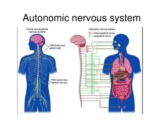
Nervous System The nervous system consists of central and peripheral parts Central nervous system Brain, spinal cord, retina & CNII Peripheral nervous system Spinal & cranial nerves (all except CNII) Autonomic nervous system (sympathetic & parasympathetic) Somatic sensory and motor nerves R G Tunstall 2013
Nervous System Outflows & Inflows Somatic motor (Skeletal muscle) Motor (Efferent) Branchio-motor (Pharyngeal arch muscle) Visceral/Autonomic motor (Organs & vessel) Somatic sensory (Sensations from somatic tissues) Sensory (Afferent) Visceral sensory (Sensations from organs & vessels) Special Visceral sensory (Taste) Spinal nerve Mixed nerve containing motor, & sensory neurons Some contain autonomic neurons R G Tunstall 2013
Autonomic Nervous System Autonomic neurons are classified as pre- and post-ganglionic Postganglionic Preganglionic Target Brainstem nucleus / Cell body in lateral horn Peripheral ganglion • These nuclei are controlled by descending reticospinal tract neurons. The latter neurons are separate to the pre/post ganglionic classification Lateral horn Autonomic (only seen T1-L2 & S2-4) R G Tunstall 2013
Sympathetic Nervous System Neurons controlling lateral horn cells (or autonomic brainstem nuclei) arise in the hypothalmus Sympathetic chain extends up to near the skull base T1 spinal nerve Sympathetic nerves exit CNS with T1 - L2 spinal nerves THERE ARE NO OTHER EXIT POINTS L2 spinal nerve Damage to the spinal cord at VERTEBRAL LEVEL L1 should not affect sympathetic innervation but will affect pelvic parasympathetic nerves Sympathetic chain extends to tip of sacrum R G Tunstall 2013
Autonomic Nervous System Sympathetic nerves emerge from the CNS only with spinal nerves T1-L2 Sympathetic chain Runs on lateral sides of vertebral bodies throughout all vertebral column regions R G Tunstall 2013
Sympathetic Nervous System The sympathetic chain communicates with spinal nerves via rami communicans Rami communicans White = preganglionic sympathetic neurons passing into chain Grey = postganglionic sympathetic neurons passing from chain into spinal nerve Don’t forget nerves such as the thoracic & cardiac splanchnic nerves emerge from the sympathetic chain Sympathetic ganglia Sympathetic chain R G Tunstall 2013
Sympathetic Nervous System The sympathetic chain communicates with spinal nerves via rami communicans Dorsal root ganglion Formed by the cell bodies of sensory neurons Spinal nerve Dorsal ramus Grey rami communicansonly No white ramus communicans seen before spinal nerve T1 or after L2 Ventral ramus Grey ramicommunicans Postganglionic sympathetic neurons passing back into spinal nerve White ramicommunicans- seen between T1-L2 only Preganglionic sympathetic neurons passing into chain Sympathetic chain ganglion R G Tunstall 2013
Sympathetic Supply to the Head Brainstem or spinal cord injury proximal to T1 cord level can affect sympathetic supply to head Reticulospinal tract neurons originate in brainstem/hypothalamus Descend to T1 & synapse with preganglionic neuron in lateral horn Exit cord with T1 spinal nerve Travel to sympathetic chain Run up chain to cervical ganglia & synapse with postganglionic neuron Postganglionic neurons enter head as plexus around internal carotid artery Supply face via external carotid artery. T1 L2 R G Tunstall 2013
Sympathetic Chain Ganglia in the Neck • There are normally x3 sympathetic chain ganglia in the neck Superior cervical C1-4 Near skull base Middle cervical C5-6 Stellate C7-T1 Near lung apex Causes of damage Pancoast tumour Tumour of skull base Lyphadenopathy Iatrogenic Trauma R G Tunstall 2013
Sympathetic Supply to the Head Sympathetic neurons are distributed in the head with cranial nerves and arteries Sympathetic neurons form a plexus around the internal carotid arterywhich join parasympathetic nerves. Distributed with arterial branches Run to pterygo-palatine ganglion from which they are distributed CN VII Distributed with branches of CN V This is the superior cervical ganglion in the neck Forms deep petrosal nerve which joins parasympathetic greater petrosal nerve R G Tunstall 2013
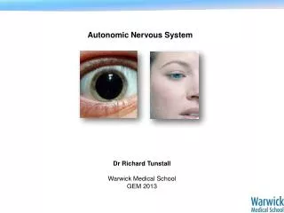
Sympathetic Supply to the Head Loss of sympathetic supply to the head leads to Horner’s syndrome R G Tunstall 2013
Sympathetic Supply to the Head Loss of sympathetic supply to the head leads to Horner’s syndrome Pupil constriction (miosis) Vasodilation Ptosis Lack of sweating (anhydrosis) Causes of damage Pancoast tumour Tumour of skull base Lyphadenopathy Iatrogenic Trauma Emerg Med J doi:10.1136/emj.2009.081679 R G Tunstall 2013
Sympathectomy The sympathetic chain or sympathetic nerves can be cut/interrupted to treat certain diseases • Hyperhidrosis Raynaud disease with tissue damage http://www.assh.org/Public/HandConditions/Pages/Cold-Hand.aspx http://bestpractice.bmj.com/best-practice/monograph/856/resources/image/bp/1.html Review the lecture video resources on Moodle R G Tunstall 2013
Sympathectomy Sympathectomy for hyperhidrosis R G Tunstall 2013
Sympathetic Nervous System Preganglionic Postganglionic Sympathetic Chain Ganglion Lateral horn cells Spinal nerves Visceral branches Arterial plexi Pre-aortic Ganglion Splanchnic nerves Adrenal Medulla Fibres also travel up and down the chain R G Tunstall 2013
Sympathetic Nervous System Chromaffin cells of adrenal medulla release adrenaline (95%) into the blood Systemic release provides a mechanism for reaching cells with no sympathetic innervation R G Tunstall 2013
Parasympathetic Outflows Parasympathetic outflow from the CNS is with x4 cranial nerves and x3 spinal nerves • Parasympathetic nuclei sit in the brainstem • Outflow with CN III, VII, IX & X Outflow with S2-4 spinal nerves Note: The S2-4 spinal nerves are formed at the L1/2 vertebral level R G Tunstall 2013
Autonomic Nervous System Parasympathetic outflow from the CNS is with x4 cranial nerves & 3 spinal nerves (conserve energy, bowel activity, urinate, defecate, erection……) CN III CN VII CN IX CN X Pelvic splanchnic nerves From S2-4 Spinal nerves R G Tunstall 2013
Parasympathetic Outflows Postganglionic parasympathetic fibres arising from cranial nerves III, VII, IX & X travel with branches of CN V Preganglionic Postganglionic Peripheral ganglion Brainstem nucleus Target Emerges with CN III, VII, IX or X Travels with CN V R G Tunstall 2013
Parasympathetic nerves in the head are hitchhikers R G Tunstall 2013
Parasympathetic Outflows Ciliary Ganglion Edinger Westphal Nucleus Eyes Pupil constriction Accommodation With CN Va With CN III Mucous membranes Structures above the maxillary teeth e.g. palate, nasal cavity, sinuses, lacrimal gland Pterygopalatine Ganglion Superior salivatory nucleus With CN VII (via greater petrosal nerve) With CN Vb Submandibular Ganglion With CN VII (via chorda tympani) Mucous membranes Structures below the the mandibular teeth e.g. sublingual, submandibular glands With CN Vc Otic Ganglion Inferior salivatory nucleus Parotid gland With CN IX With CN Vc (via lesser petrosal nerve) Organ-specific ganglia Dorsal nucleus of vagus With CN X NOTE: POSTGANGLIONIC FIBRES DISTRIBUTED WITH CN V BRANCHES R G Tunstall 2013
Parasympathetic Outflows Ciliary Ganglion (CN III) Pterygopalatine Ganglion (CN VII) R G Tunstall 2013
Parasympathetic Outflows Submandibular (CN VII supplied) & Otic Ganglia (CNIX supplied) Parasympathetic nerves to submandibular & sublingual glands (CN VII) CN V Parasympathetic nerves to parotid gland (CN IX) Otic ganglion Submandibular ganglion R G Tunstall 2013
Parasympathetic Outflows The pelvic splanchnic nerves emerge from the S2-4 spinal nerves Pelvic splanchnic nerves Branch from S2-4spinal nerves Autonomic nerve plexi Form around most pelvic organs R G Tunstall 2013
Clinical Problems • Loss of parasympathetic pupillary innervation • Frey / Frey-Baillarger syndrome No pupil light reflex (direct or consensual) Unopposed sympathetic action Gustatory sweating of the face following parotid surgery/injury Parasympathetic nerves regrow to innervate the sweat glands muscarinic receptors R G Tunstall 2013
Autonomic Innervation There are two principal transmitters in the peripheral nervous system Noradrenaline (norepinephrine) Acetylcholine R G Tunstall 2013
Autonomic Innervation There are two principal transmitters in the peripheral nervous system CNS ACh Somatic efferent NA ACh Sympathetic ACh ACh Parasympathetic R G Tunstall 2013
Autonomic Innervation The receptors for acetylcholine and noradrenaline CNS ACh Somatic efferent Nicotinic ACh NA Sympathetic a or b Nicotinic ACh ACh Parasympathetic Muscarinic Nicotinic R G Tunstall 2013
Autonomic Innervation Different receptors bring about different effects …………..even the same receptor can have different effects R G Tunstall 2013
Autonomic Innervation R G Tunstall 2013
Autonomic Innervation Muscarinic receptors are membrane-bound receptors working via G-proteins R G Tunstall 2013