Download
1 / 68
730 likes | 1.86k Vues
CONTACT DERMATITIS. Oktay Taşkapan,MD. Ekzema-Dermatit. Polimorf inflamasyon paterni Kontakt dermatit: Derinin ekzematöz reaksiyonuyla kendini gösteren inflamatuar bir intolerans (irritasyon / allerji) İKD daha sık, (etken belirlenmezse) AKD’nin prognozu daha kötü

E N D
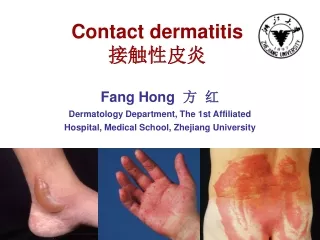
CONTACTDERMATITIS Oktay Taşkapan,MD
Ekzema-Dermatit Polimorf inflamasyon paterni Kontakt dermatit: Derinin ekzematöz reaksiyonuyla kendini gösteren inflamatuar bir intolerans (irritasyon / allerji) İKD daha sık, (etken belirlenmezse) AKD’nin prognozu daha kötü KD insidansı %0,79 ve en az bir allerjene duyarlanma %15-16 * KD, dermatolojik konsültasyonların % 4-7’sinde saptanır *Brasch J ve ark. JDDG 2007.
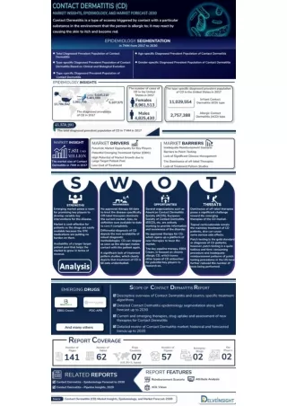
Contact Dermatitis • A spectrum of inflammatory skin reactions induced by exposure to external substances • Contact dermatitis represents the vast majority (79–90% annually) of skin-related occupational complaints • A common skin problem. Nearly eight million physician visits per year (2000, USA) • More than 85 000 chemicals, almost 3000 of the may act as contact allergens • When avoidance and appropriate treatment isn’t achieved, the condition may become chronic and lead to a major impairment in quality of life • It is a major health concern for patients and has a major impact on the economy
A lifetime incidence of “hand eczema” is 17 % (Danish studies) • For contact dermatitis without further etiologic definition, i.e. irritant and allergic combined, an incidence of 7.9 per 1000 population per year was observed • Incidences in selected professions are significantly higher
In contrast to ACD, there is no specific recognition of a foreign substance in ICD, but a reaction of the immune system towards skin damage • Even in ICD, inflammatory mediators released in the framework of a complex network by cells of the epidermis and dermis as well of the immune system play a central role • Routine tests such as histology do not allow differential diagnosis between ICD and ACD.
Acute stage: Erythematous papules, edema, and vesiculation • Subacute stage: The vesicles rupture oozing and eroded skin crusting and scaling • As the papulovesicular lesions disappear, ACD enters into chronic stage (lichenification, further scaling, fissuring and pigmentation) • Secondary bacterial infections may occur
Irritant contact dermatitis (ICD) • Allergic contact dermatitis (ACD) • Photocontact dermatitis (phototoxic / photoallergic)
Irritant contact dermatitis: lesions limited to sites of contact, spectrum ranges from erythema to necrosis, clinical picture dependent on acuteness and agent • Allergic contact dermatitis: spread from primary site of contact is typical. Location and configuration of lesions can be indicative of causative agent • Airborne contact dermatitis: dermatitis on exposed skin without overt allergen contact (wall paints, flowering plants, others) • Photo contact dermatitis: occurs primarily on light-exposed skin. Previous contact not necessary for irritants, but definitely for photoallergens • Asteatotic dermatitis: dry, “fissuring” skin with red fissures especially in aged and overly stressed skin (wrong skin care, excessive washing).
Airborne contact dermatitis (due to parthenium hysterophorus)
Allerjik kontakt dermatit • Antijene özgüllük gösteren efektör T hücrelerinin rol oynadığı bir deri inflamasyonu • Yaşam boyu prevalansı yaklaşık %15 (Brasch J ve ark. JDDG 2007) • Kaşıntı, vezikülasyon ve papül oluşumu daha belirgin • İlk ortaya çıkan lezyonlar, temas bölgesinde sınırlıdır, ancak uzak yayılım da gelişebilir
Allergic contact dermatitis can occur anywhere on the body • There may be extreme pruritus and the lesions may spread beyond the areas of initial contact • The appearance of the lesion in ACD often corresponds to the stage • Edema predominates if areas of loose tissue, such as eyelids or genitalia, are affected • The vesicles may coalesce into bullae
Yama testi: IPPD: (++) Yama testi: Potasyum dikromat, kolofoni, karba miks ve tiuram miks (++)
İrritan kontakt dermatit • Derinin irritanlara karşı geliştirdiği non-immünolojik / non-spesifik reaksiyon • Genellikle yüksek dozda irritan maddeyle karşılaşan hemen herkeste, özellikle atopik bireylerde görülür • Kuaförler, besin sektöründe çalışanlar ve sağlık çalışanlarında İKD gelişme olasılığı yüksektir • Doğrudan doku yıkımıyla birkaç dakika ile birkaç saat içinde ortaya çıkar, genellikle keskin sınırlıdır • Akut evrede kaşıntıdan çok yanma, batma, acıma ve ağrı belirgindir • Kronik evrede hiperkeratoz ve fissürler görülür
Akut İKD (dilüe edilmemiş alkali temizleyiciye bağlı) Akut İKD (Monokloroasetik asite bağlı) İKD (bromoasetik aside bağlı) Akut İKD (diklofenak jel kullanımına bağlı) Akut İKD (ağdaya bağlı) Yüzde HTİKD (taş tozuna bağlı)
İrritan el ekzeması Kronik İKD (temizlik işçisi) Kronik İKD (metal işçisi) İKD (Aşırı el yıkamaya bağlı) Kronik İKD (hemşire) Kronik İKD (basımevi işçisi) Kronik İKD Kronik İKD Kronik İKD (numuler tip)
A specific diagnostic test to identify irritant contact dermatitis is not available • The diagnosis of irritant contact dermatitis is made on the basis of history and clinical findings after excluding a causative contact sensitization, and can indirectly be confirmed by successive healing after stopping exposition to the noxious agent • DD: Atopic dermatitis, fungal infections, cutaneous T-cell lymphoma or special forms of psoriasis
AKD’de Lezyonların yerleşimi ve tanısal ipuçları Lee PW ve ark. Curr Opin Pediatr 2009.
CD4+ or CD8+ T cells play critical roles in ACD • Langerhans cells, NKT cells, NK cells, B cells, and Treg cells • Although most environmental agents are too large to penetrate the skin layers, some are of sufficiently low molecular weight to penetrate the stratum corneum • Hapten exposure duration and the host immune response are crucial components in the development of ACD.
ACD: Histopathology • A pattern of subacute chronic dermatitis or acute dermatitis may be seen • The inflammatory infiltrate in the dermis predominately contains lymphocytes and other mononuclear cells • Epidermal edema (ie, spongiosis and microvesicle formation) may be seen • Chronic stage: Thickening of the epidermis (acanthosis) with hyperkeratosis and parakeratosis
Epidermal spongiosis and edema Epidermotropism of mononuclear cells Dermal inflammatory cellular infiltrate
ICD Asteatotic eczema Atopic dermatitis Seborrheic dermatitis Nummular dermatitis Dyshidrotic dermatitis Photocontact dermatitis Intertrigo Tinea corporis Tinea cruris / pedis Dermatomyositis Lichen simplex chronicus Cutaneous T-cell lymphoma Perioral dermatitis Stasis dermatitis Cellulitis ACD: Differential diagnosis
Differential diagnosis of acute ACD • Acute ICD • Erysipelas • Acute SLE • Dermatomyositis • Angioedema • Herpes zoster (early phase)
Severe edema of the face 2 days after dyeing thehair at home (a). The patient was referred by the emergencyphysician as erysipelas because she had slight fever, nausea,and lymphadenopathy. Close inspection revealed eczematouslesions at the hairline and on the scalp (b).
Metals • Nickelis the most common cause of ACD in women • High-nickel content jewelry is a redisposing factor • Earpiercing is considered to be the principal inducer of nickel CD • Hand eczema in nickel sensitive patientsis often of the dyshidrotic type and may be aggravated bynickel ingestion • Chromateis the most common contact allergen in men • Sensitization to it is usually occupational. • Occupationalexposure is most frequent in construction workers whohandle cement • Other common sources are chrome-tannedleather, bleaching agents, paints, and printing solutions.
Cosmetics and skin care products • Compulsory ingredient labeling of cosmetic products has greatly facilitated the diagnosis andtreatment of cosmetic contact dermatitis • Positive patchtests are found most frequently to preservatives, perfumes,active or category-specific ingredients, excipients/emulsifiers and sunscreens • The relevance of thepositive patch tests is confirmed if the contact dermatitisdisappears upon discontinuation of the use of the product • Most allergic reactions are caused by cosmetics that remainon the skin: “stay-on” or “leave-on” products
Dermatitis from clothes and shoes • Contact dermatitis to clothes is usually located in the axillae • Clothing dermatitisfrom formaldehyde is rare nowadays. • Textile dye dermatitisis usually related to disperse dyes • Leather articlescontain several substances that may cause ACD: chrome, adhesives (paratertiary butyl phenol formaldehyde resin), and dyes • Anumber of accelerators and antioxidants used inthe production of synthetic rubber may also cause contact dermatitis.
Drug dermatitis • Drug dermatitis may be elicited by the active ingredient ofa topical drug, by the vehicle or by a preservative • Contactsensitization to antibiotics, antiseptics, and anesthetics isrelatively frequent, especially in leg ulcer patients • ACDfrom topical corticosteroids has been reported with increasingfrequency • Systemic application of a drug towhich an individual has been sensitized by a previouscutaneous exposure may cause systemic contact dermatitis
History and clinical findings are guides to the diagnosis • History should include questions on the development of the disease, exposition to allergens and causal relationships and be renewed after the results of patch testing • Exposition and clinical findings lead to the suspicion that the dermatitis might be caused by contact with an exogenous agent • The diagnosis of ACD is made by demonstrating the causative contact sensitization in patch testing • In vitromethods in diagnosing contact allergy have not been validated
Patch testing • Patch testing is cost effective and reduces the cost of therapy in patients with severe ACD • At the time of patch testing, the patient's dermatitis must be under excellent clinical control • The dermatitis cannot be severe or acute, as this may result in the false-positive, ‘angry back’ reaction • Ptients should not be taking high-dose systemic corticosteroids (low dose, up to 10–20 mg/day, may be acceptable) • It is also advisable to avoid the application of potent topical corticosteroids to the patch test sites
Patch testing • Patch testing is required to confirm the diagnosis of ACD and to make a differential diagnosis • Patch testing must be performed by health care providers trained in the proper technique • Patch testing procedure: Small amounts of appropriate labeled dilutions of chemicals are applied to the skin and occluded for 2 days • The patch test must be read not only at 48 hours, when the patch tests customarily are removed, but again between 72-96 hours and 1 week following initial application