
Influence of Disability on Trunk Muscle Activity in Chronic Low Back Pain
This study investigates how self-reported disability levels in chronic low back pain patients affect trunk muscle activity during maximum effort isometric trunk exertions. The research examines muscle activation patterns in different trunk movements. Results show significant differences in muscle activity based on disability levels.

Influence of Disability on Trunk Muscle Activity in Chronic Low Back Pain
E N D
Presentation Transcript
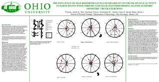
A B A side view extension B side view flexion C top view extension D top view right rotation E top view left rotation F top view flexion G top view right side bending H top view left side bending C F D G Effort 1. Extension 2. Right rotation 3. Left rotation 4. Flexion 5. Right side bending 6. Left side bending E H THE INFLUENCE OF SELF-REPORTED LEVELS OF DISABILITY ON TRUNK MUSCLE ACTIVITY IN PARTICIPANTS WITH CHRONIC LOW BACK PAIN PERFORMING MAXIMUM EFFORT ISOMETRIC TRUNK EXERTIONS Thomas, James S.; Sha, Daohang; France, Christopher R*.; Swank, Kevin A; Vander Wiele, Nicole J. School of Physical Therapy, *Department of Psychology, Ohio University, Athens, OH Introduction Many theories exist concerning the effect of low back pain on trunk muscle activation. In a pain-spasm-pain model during maximal concentric contractions it is predicted that individuals with and without back pain will have equal muscleactivation. In contrast in a pain-adaptation model it is predicted that individuals with pain will have less EMG activity in the agonist muscle groups and increased activation of the antagonists in an effort to stabilize and support the spine. The purpose of this study was to examine the influence of self rated disability in chronic low back pain subjects on trunk muscle activity during maximal voluntary isometric trunk exertions. Methods Prior to the start of the maximal exertions, 20 participants with chronic low back pain completed the Roland Morris Disability Questionnaire (RMDQ). The participants were positioned in an apparatus so that they were in a half-kneeling position with the lumbar spine in a neutral position and the pelvis immobilized. A cable was run from a thoracic harness to a load cell fixed to the structure. Motion of the lumbar spine was constrained to a neutral posture. Figure 1 illustrates the direction of trunk movement and the load cell and cable set up for each of the tests of maximal exertion. Participants were instructed to cross their arms over their chest and pull as hard as they could against the cable. Participants completed two trials of maximal effort exertion in flexion, extension, and rotation (left & right). Participants were instructed to maintain their maximal effort for five seconds. Trunk force was determined from the load cell connected in series to the thoracic harness. The load cell data were sampled at 1000 Hz on the same system as the EMG data. Muscle activity of the left and right rectus abdominis, external oblique, internal oblique, iliocostalis lumborum, and multifidis were recorded at 1000 Hz using a 16 channel Delsys Bagnoli system. The EMG data were rectified and low pass filtered using a 4th order zero lag butterworth filter with a cut-off frequency of 2.5Hz. The EMG data for each muscle were averaged from the point of maximal force on the load cell until the end of the 5 second exertion. Data Analysis To analyze these data, first, the chronic low back pain subjects were divided by a median split on the RMDQ. To test for group differences (high RMDQ or low RMDQ) on trunk muscle activity, separate ANCOVAs were used to analyze averaged peak EMG of each trunk muscle while controlling for differences in force generated (i.e. maximum force on load cell).Results During maximal trunk flexion exertions (Fig 2), the high RMDQ group had significantly less EMG activity in six out of eight trunk flexors (p<.05). The graphical representation illustrates that neither group used co-contraction of the trunk extensors. For trunk extension exertions (Fig 3), compared to the low RMDQ group, the high RMDQ group had significantly less EMG activity in three out of four trunk extensors (p<.05), even when controlling for the magnitude of force generated. Both groups demonstrated mild co-contraction but only in the internal oblique muscle. In right rotation exertions (Fig 4), the high RMDQ group had less EMG activity in left external oblique, right internal oblique, both erector spinae, and the left multifidis (p<.05). Both groups used a similar activation pattern relying on the right erector spinae and internal oblique as well as the left external oblique to generate the movement. In left rotation exertions (Fig 5), the high RMDQ group had less EMG activity in all trunk flexors except the left internal oblique (p<.05). Once again the two groups used similar patterns of activation, however those with high RMDQ scores used less force during muscle contractions. In right side bending exertions (Fig 6), the high RMDQ group had significantly less EMG activity in all five of the R sided muscles (p<.05). In left side bending exertions (Fig 7), the high RMDQ group used significantly less EMG activity in four of the five left sided muscles (p<.05). During side bending in either direction both groups demonstrated co-contraction of the opposite side internal and external oblique muscles. Conclusions Even when controlling for force generated, individuals with high levels of self reported disability use less trunk muscle activity compared to individuals with low levels of disability. This research was supported by The National Institutes of Health Grant R01-HD045512 to J.S. Thomas Figure 1: A-H illustrate the set-up of the load cell and thoracic harness for each of the maximal muscle tests Figure 2: Flexion Figure 6: Right Side Bend Figure 4: Right Rotation LOW RMDQ HIGH RMDQ Figure 3: Extension Figure 7: Left Side Bend Figure 5: Left Rotation


